Compare the different methods of neuroimaging. How are these methods alike and different?According to Argosy (2008) some different methods of neuroimaging include X-rays, CT, MRI, PET, fMRI, and EEG.
In x-rays, a beam is passed through the object or person onto a photographic plate. Pinel (2009) This beam is absorbed differently by different materials. Usually, hard materials such as metal and bone absorb it quite well and really show up on the x-ray photograph. However, other materials, such as clothing or soft tissue do not show up as well, because they do not absorb as well.
A CT, Computed Tomography, scan uses x-rays in rotation to take and combine many individual x-rays around the brain. Then the machine is moved along the axis of the patients body. Pinel (2009) and adventually the information is combined to make a 3-D photo of the brain.
Next, an MRI, or Magnetic Resonance imaging, which uses magnets and measures waves produced by these magnetic fields.
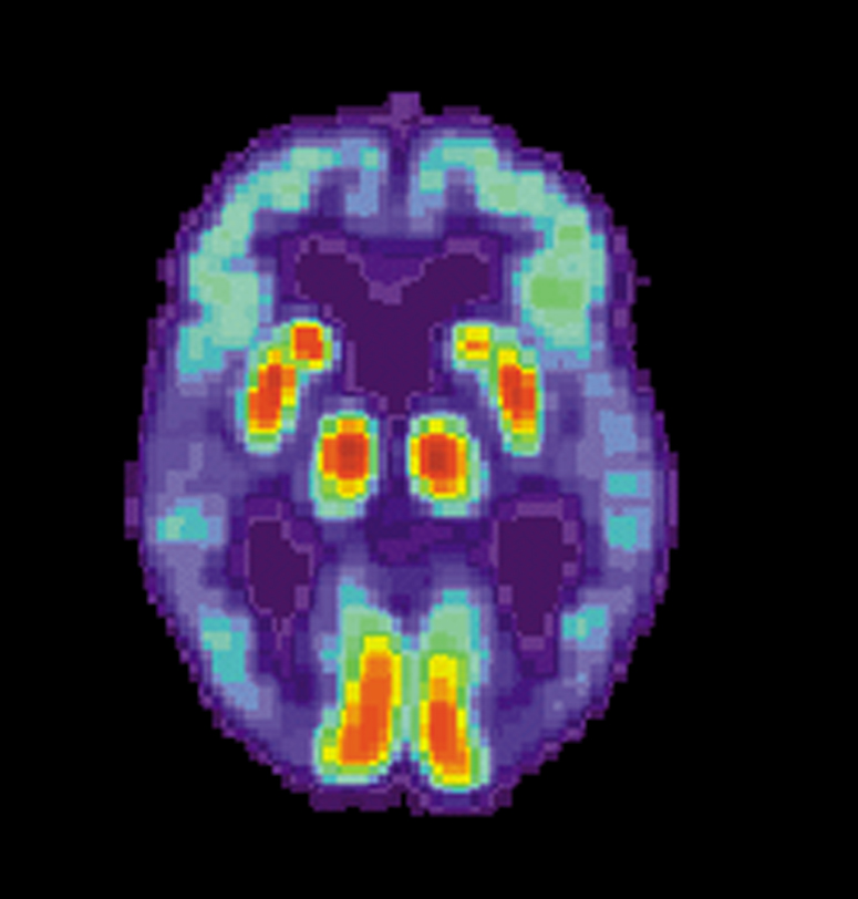 English: PET scan of a human brain with Alzheimer'...
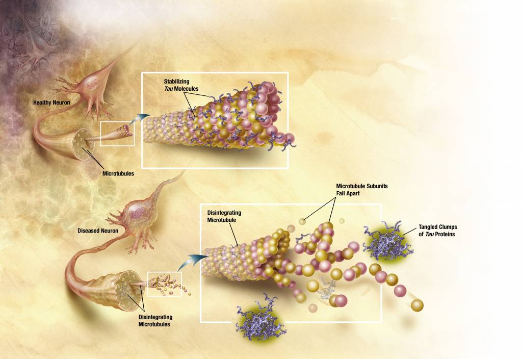 English: Diagram of how microtubules desintegrate ...
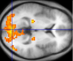 A fMRI scan showing regions of activation in orang...
Pinel (2009). The MRI image is ideal for showing special locations in the brain structure. Pinel (2009). I was recently exposed to this with a relative of mine who was sick and we feared had a stroke. They first took a CT scan, but, a few days later wanted to take an MRI. I asked the doctor why this was, and he said that although a CT scan should pick up if there was a big pool of blood, the MRI machine would pick up blockage images and they could use it to scan in finer detail. Thankfully, his test results came back negative for stroke, but it was a good educational experience on how each procedure can produce unique but useful sets of information.
PET, or Positron Emission Tomography, differs from the previous mentioned scans in that it...
Comparison?
Good information was provided but I would have liked to see more comparison as the introductory statement stated it would describe how they are alike and different. Instead, paper describes each topic without much detail. Some good info in there though!
2 out of 2 people found this comment useful.