Outline
Spinal Cord, Spinal Nerves, Somatic Reflexes-Chapter 13
Reflexes- they are involuntary stereotyped responses to stimuli, they involve the brain, spinal cord, and peripheral nerves
Spinal cord- cylinder nervous tissue that begins at the foramen magnum and passes through the vertebral canal as far as the inferior margin of the first lumbar vertebrae (L1), 18 inches long and ý inches wide
Anterior Median Fissure- in the front, deeper
Posterior Median Sulcus- in the back, shallow
âªTwo Enlargements for Limbs: (up and down below)
1. Cervical Enlargement- in the inferior cervical region, the spinal cord gives rise to the nerves of the upper limbs, up
2. Lumbar Enlargement- in the lumbosacral region there are nerves to the pelvic region and lower limbs arise, below
Medullary Cone- the spinal cord becomes more tapered
âªMeninges- the spinal cord and brain are enclosed in three fibrous membranes, specialized membranes that surround the spinal cord
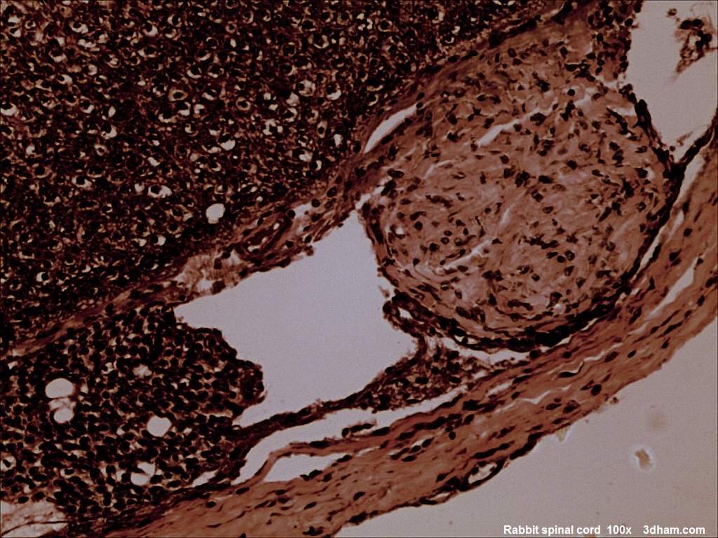 English: Rabbit spinal cord.
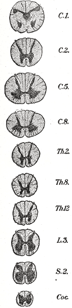 Cross-sections of the spinal cord at varying level...
 Cross-section through the spinal cord at the mid-t...
A. shock absorption
B. physical stability
C. blood vessels that branch within these layers to provide oxygen and nutrients
1. Dura Mater- outer most layer, forms a loose fitting sleeve called the dural sheath around the spinal cord, though and fiborous, it will fuse with the periosteum of the occipital bone around the formamus magnum
Coccygeal Ligament- inferiorly the dura mater will blend with the filum terminal, longitudinal support
-horizontally there is stability by fusing with connective tissue of the spinal nerve such that the dura mater will extend out of the IVF
Epidural Space- the space between the sheath and the vertebrae bone, the space between the dura and the wall of the vertebral canal, it has loose connective tissue, blood vessels and adipose tissue
2. Arachnoid Mater- adheres to the dural sheath, it consists of a simple squamous epithelium, the arachnoid membrane
Subarchnoid Space- a loose...
Anatomy&physiology
100% Summary did help me alot.
0 out of 1 people found this comment useful.