A. Cell Theory
1. All organisms, both unicellular and multicellular, are made up of cells.
2. Cells are the smallest units of living matter and structural and functional units of all organisms.
3. In 1830's, Mathias Schleiden (plants) and Theodore Schwann (animals) declared organisms were made of cells.
4. Cells are capable of self-reproduction; Rudolf Virchow declared cells come only from preexisting cells.
B. Cell Size
1. Cells range in size from a frog's egg (one millimeter) down to one micrometer.
2. Cells need surface area of plasma membrane large enough to adequately exchange materials.
3. Surface-area-to-volume ratio requires that cells be small.
a. As cells get larger in volume, relative surface area actually decreases.
b. Limits how large actively metabolizing cells can become.
c. Cells needing greater surface area use modifications such as folding, microvilli, etc.
C. Microscopy of Today
1. Bright-field microscope uses light rays focused by glass lenses.
 Bacterial cells of Staphylococcus aureus, which is...
 Cell biology
 Pollen from a variety of common plants: sunflower ...
2. Transmission electron microscope (TEM) uses electrons passing through specimen; focused by magnets.
3. Scanning electron microscope (SEM) uses electrons scanned across metal-coated specimen.
4. Magnification is function of wavelengths; shorter wavelengths of electrons allow greater magnification.
5. Resolution is minimum distance between two objects before they are seen as one larger object.
6. Immunofluorescence microscopy uses fluorescent antibodies to review proteins in cells.
7. Confocal microscopy uses laser beam to focus on shallow plane; forms series of optical sections.
8. Video-enhanced contrast microscopy accentuates the light and dark regions and may use a computer
to contrast regions with false colors.
9. Bright-field, phase contrast, differential interference and darkfield are different types of light microscopy
that improve ability to see various features.
 Cell Biology
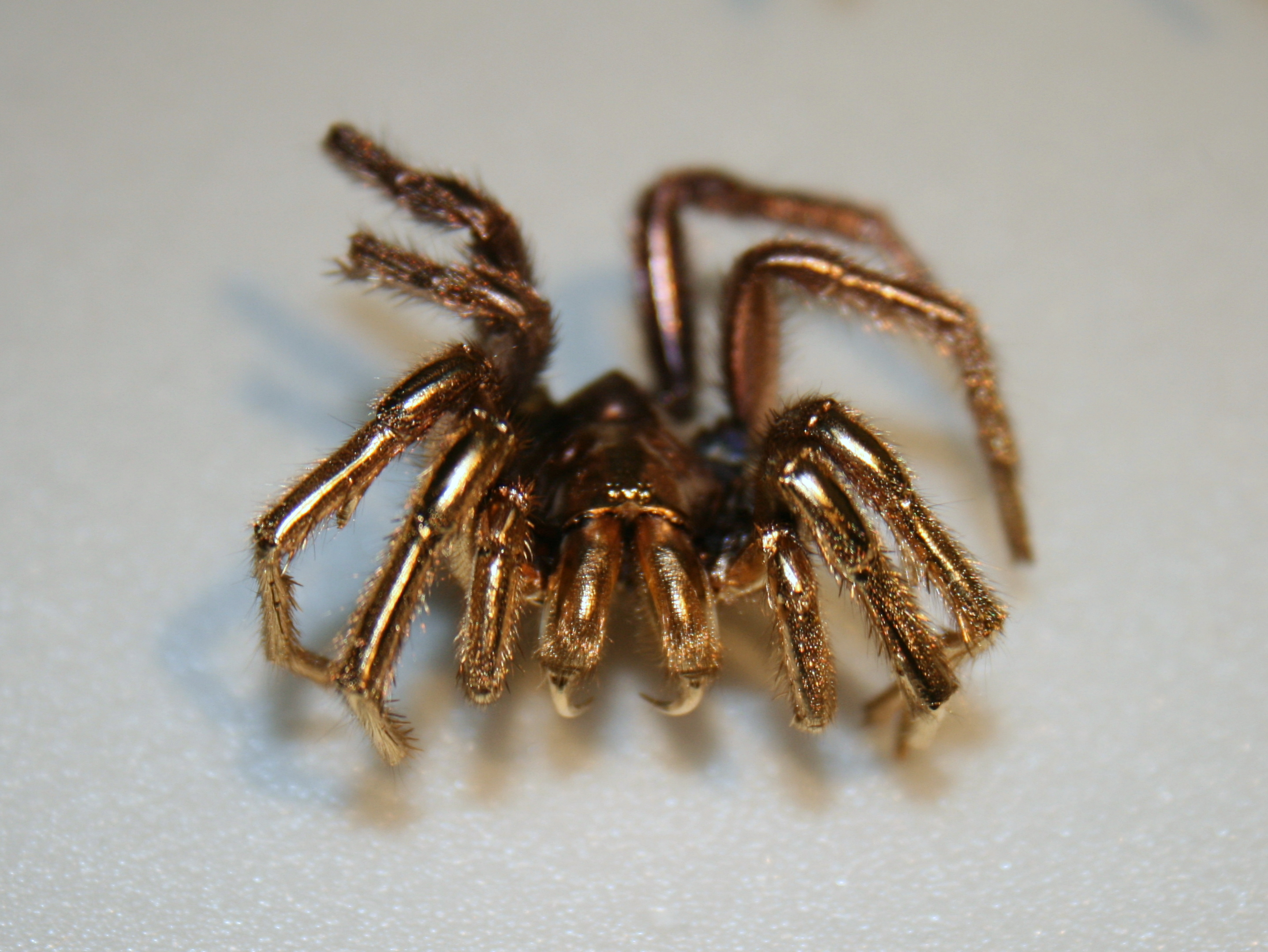 English: A spider coated in gold to prepare it as ...
 "Scanning Electron Microscope"