Overview:
Computed Tomography (CT) imaging, also known as "CAT scanning" (Computed Axial Tomography) is a invaluable medical diagnostic imaging tool, which Uses a combination of X-rays and computer technology. A series of X-ray beams from many different angles are used to create cross-sectional images or "slices" of the patient's body. These images are assembled in a computer into a three- dimensional picture that can display organs, bones, and tissues in great detail.
The CT scanner consists of a large donut-shaped machine and an X-ray table. The Patient will lie on the table and be slowly moved into the large opening as a series of pictures are taken. Depending on which area of the body being scanned a "contrast" may be given. The contrast agent (orally, rectally or via injection) also known as a "dye" improves the visibility of structures within the body and filtered out of the bloodstream naturally by kidneys and the liver.
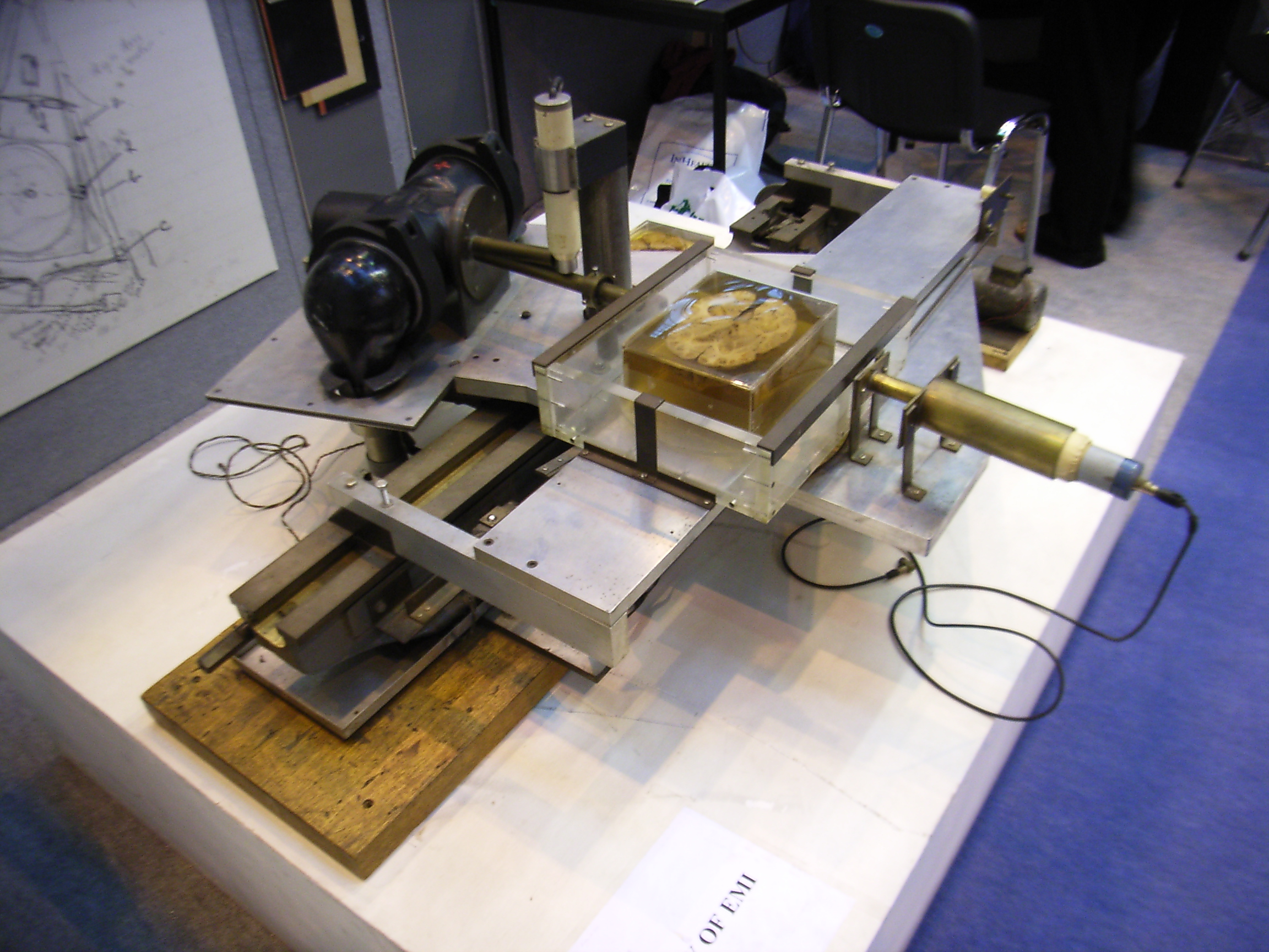 The prototype CT scanner
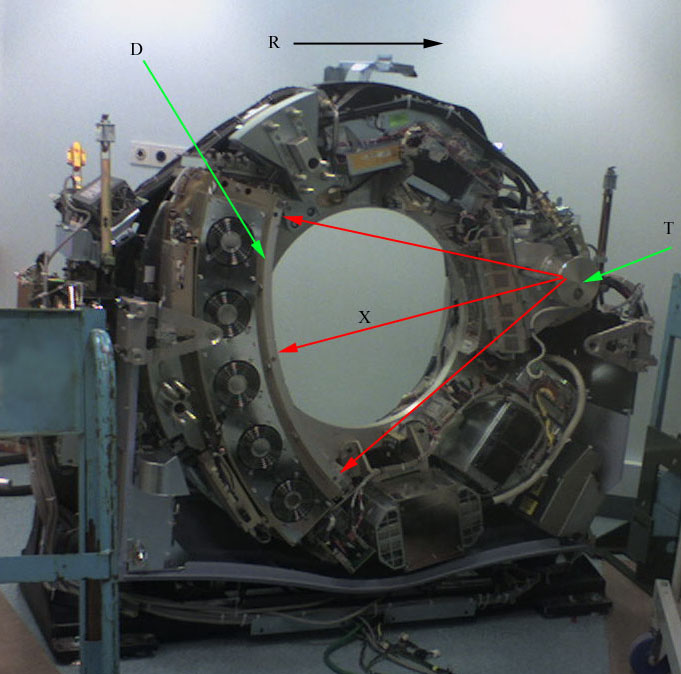 CT scanner with cover removed to show the principl...
 X-ray bath
CT scans are performed to analyse the internal structures of various parts of the body. This includes the head, where traumatic injuries occur, such as blood clots or skull fractures. Tumours and infections can be identified. In the spine, the bony structure of the vertebrae can be accurately defined, as can the makeup of the intervertebral discs and spinal cord.
History And Development
CT was invented in 1972 by British engineer Godfrey Hounsfield of EMI Laboratories, England, and independently by South African born physicist Allan Cormack of Tufts University, Massachusetts. Hounsfield was later awarded the Nobel Peace Prize and honoured with Knighthood in England for his contributions to medicine and science.
The first clinical CT scanners were installed between 1974 and 1976. The original systems were dedicated to head imaging only, but "whole body" systems with larger patient openings became available in 1976. CT...