X-rays, the savior of hospitals, the scientific interest of many. In October 1895 a German physicist named Wilhelm Konrad Roentgen stood round a device called a Crooke's tube, trying to find out more about it. He had covered the tube with black paper, and in a dark laboratory room, he noticed an eerie green glow appeared when the tube was turned on. No matter how many pieces of cardboard he placed in front of the tube, the glow continued. A photographic plate that was lying near the tube was blackened, and from this he could tell that some type of invisible rays must be coming out of the Crooke's tube. Roentgen named these 'invisible rays' x-rays, x being the unknown.
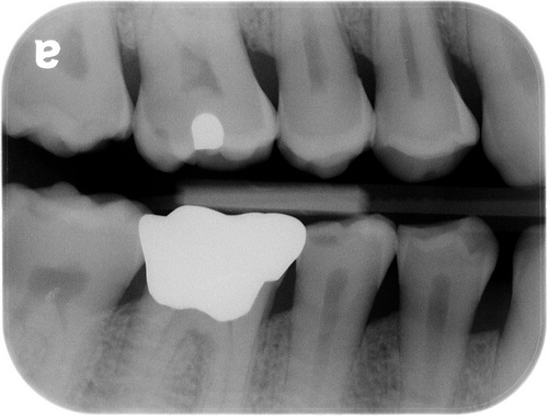
One place x-rays are being used in society is in dental clinics and hospitals. An x-ray picture, or a radiograph is used to take pictures of limbs, bones and organs, they go through the tissue but are stopped by the bones.
 x-ray
x-ray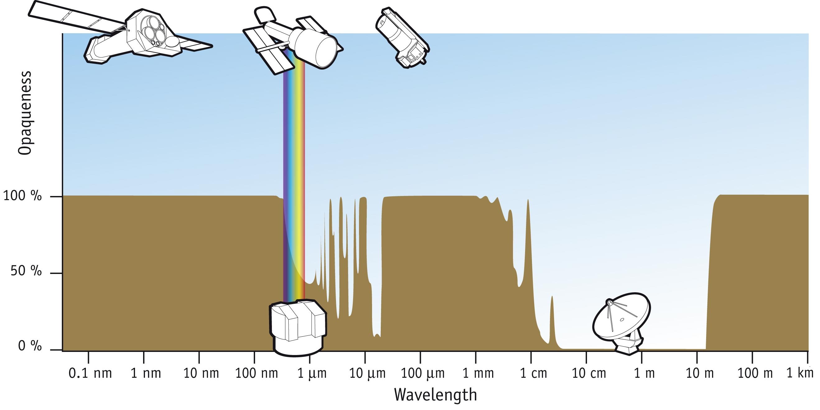 Gamma ray absorbation in the atmosphere
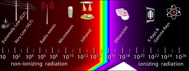 OSHA radiation spectrum
In a hospital, a radiographer will stand behind a screen, while the patient being x-rayed stands between an x-ray tube and photographic film, and the rays will pass through the body on to the film. Though x-rays allow doctors to see through the skin that beforehand could only be seen by cutting a person up, too much x-radiation can be deadly and lead to cancer. The risk of getting cancer from x-rays is .01%; even so the patient and the radiographer must take special precautions to be protected from these harmful rays. The patient will have everything but the area being x-rayed covered by lead, and the radiographer may wear a lead suit, and/or have a lead screen in front of him. Lead is used because x-rays are almost completely stopped by this metal. X-rays travel at...