OAC BIOLOGY LAB #1 CELL FORM AND FUNCTION LAB HOME/LINKS The design of organisms, organs, tissues, cells and even biochemicals is directly related to the function that they perform. This can be seen from the predatory design of a tiger down to the molecular design of stomach enzymes. To make sure you have a sense of this relationship, start with something easy. In a sentence, describe how one aspect of the form of a tiger fits its role as a predator in the environment.
ACTIVITY LEARNING OUTCOME: By conducting this lab it will allow you to study the relationship between form and function at a cellular level. First you will examine the cells outlined in the table below. This table also lists the function of the cell and the name of the slide(s) upon which it can be found. Once you have collected this data, you will propose logical connections between the form of the cell and its function.
 Cell biology
 English: Schematic of gram-positive cell wall show...
 Cell Biology
During your lab time 1. Prepare a proper biological drawing for any four Animal cells and any four Plant cells listed below.
2. Your drawing will be carefully copies the form of the cell - do not generalize or simplify the form of the cell. You only need to draw one cell of each type.
3. Estimate the size of each cell and indicate this on your diagram.
4. Have your diagram signed by your instructor to assure you have an accurate view of the cell.
5. Details of proper drawing methods and sizing technique can be found below using this link. BIOLOGICAL DIAGRAMS CELL NAME FUNCTION SLIDE NAME(S) ANIMAL CELLS Epidermis Covering and Protection Frog Skin Nerve Conduct Nerve Impulses Giant Multipolar Neuron Blood Conduction of Oxygen Frog, Bird, or Fish Blood Sperm Transportation of Genetic Information Rat, Bull or Human Sperm Lung Allow for Exchange of Gases Human Lung PLANT CELLS Pollen Carries Genetic Information to the Egg Pine (Pinus) Pollen Xylem Rigid Support and Water Conduction Pine Wood (Stem) Pith Storage of Minerals Tilia (Centre of Stem) Cortex Photosynthesis Ranunculus (Perimeter of Stem) Spores Carried by wind to germination site where it grows into a new individual Equisetum Spores During your study time, write a short note for each cell that suggests how its form (size, shape, arrangement, thickness of wall, etc.) could help it perform its function effectively. These will be conjectures on your part.
Pinus Pollen Grain~ From my drawings it looks as if the middle contains DNA strands or some type of genetic material and it is also well protected by a mass of green surrounding it which might help the cell carry and protect the genetic information to the egg. It also has a very thick cell membrane surrounding it which protects the information inside. It is also quite a big cell which allows it to be a stronger more efficient carrier. Elaters are there for wings and elevation.
Pith Tilia~ This cell looks pretty much like an octagon shaped bubble and the shape looks like it could grow in size which would make it suiting to store minerals, looks somewhat expandable and like a vacuole. The thick cell wall keeps the nutrients inside even though it is thin in comparison to other cells since the cell is already protecte3d in the middle of a tree. The shape makes it only able to be arranged in a certain manner which makes it more compact.
Bull Sperm Smear~ The sperm has a large area at the top which carries the information safely as well as a tail or flagella for rapid movement. Its small compact shape allows the cell to move more rapidly and squeeze through tight places in order to transport the genetic information effectively.
Frog Blood Smear~ Blood cells carry oxygen throughout the body so they are small and transportable and the cell wall which surrounds them is thin to carry the oxygen inside with no leaks but so that they can exchange oxygen easier. These cells are easily arranged and portable. Smooth shape so it can slide through the capillary walls. Arranged separately but close together so they can go in their own directions but a lot close together carries more oxygen. Large cytoplasm has hemoglobin. Lung Cell~ These cells are arranged very closely together so that they can perform their function of exchanging oxygen. The cells are the perfect size to make this exchange a fast process and they are also thick enough"æ"æ"æ"æ"æ"æ"æ"æ"æ.
Giant Multipolar Neurons Smear~ The shape of the cell shows that it reaches to many different areas and the intertwining strands could help in the conducting of nerve impulses since they are all over each other. This cell is quit large which might be because it has such a large job o do in carrying out impulses. The most important feature of this cell would probably be its shape as it reaches to everywhere which is so important because the nerve impulses need to reach all over the body.
Equisetum Spores~ A small cell which could make it easier to be carried to germination site. Since they are flat they are carried more easier and the thick mass surrounding it helps it to stay intact as it is being carried. Seed which is one cell big. It has this form because it needs a thick cell wall for protection since it travels so much. They also have elaters to carry and to hook them into the ground.
Cortex Cell~ The many chloroplasts in the cell are held in by a thick cell membrane. The shape allows for the cells to arrange together quite easily and therefore have more of a surface area so that photosynthesis will take place more effectively and faster. Chloroplasts are visible in this cell. Found on the outside of the stem so that they can catch sunlight. Have a large central vacuole.
During our class discussion, we will review the form of the cell and its size. Then we will collect individual suggestions of how its form helps it perform its function effectively. This will give you an opportunity to add to you answers.
BIOLOGICAL DRAWINGS Accurate drawings are essential to the study of biology because they allow you to become familiar with the object being studied through detailed observation and they give you a useful record to review.
Helpful hints for making better drawings: 1. Use high quality paper, a sharp, hard pencil (black only), and use an eraser to correct minor errors.
2. Observe the object carefully before you start to draw. If it is on the microscope, view it from different angles and with different lighting.
3. Sketch a large (at least half a page), faint, outline first to get the proportions set, and then go back and add the details all the while copying directly from the specimen instead of being "creative"�.
4. Be sure to improve on the view by eliminating all unnecessary details (broken cells, air bubbles, other specimens etc) 5. Add shading, labels, notes, details of size(see note below), title and date 6. Have your drawing signed by your instructor before you put the specimen away ESTIMATING THE SIZE OF AN MICROSCOPIC OBJECT 1. Move the slide around until the object is lined up next to the calibrated arrow seen in the eyepiece (an enlarged view is seen below).
2. Count the number of divisions on the scale of the arrow that object spans.
3. Convert this count into MICRONS using the values given in the chart seen below.
4. Be careful to use the values in accordance with which power (magnification) you are using.
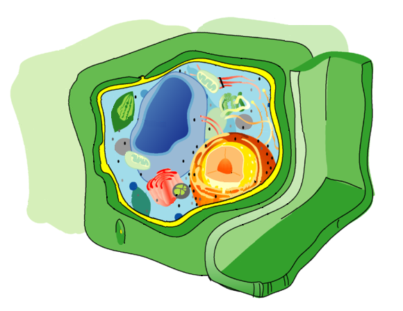 Diagram of the plant cell, with the cell wall in g...
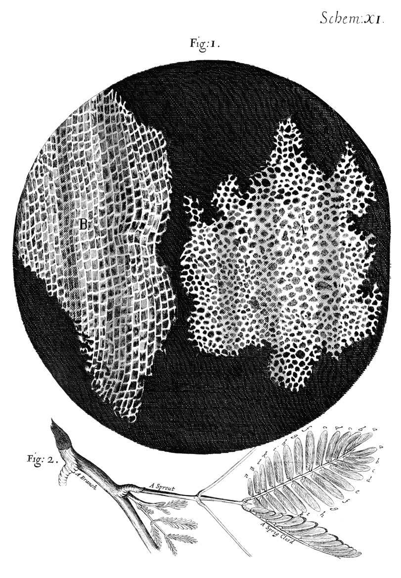 The cork described in "Micrographia" by Robert Hoo...
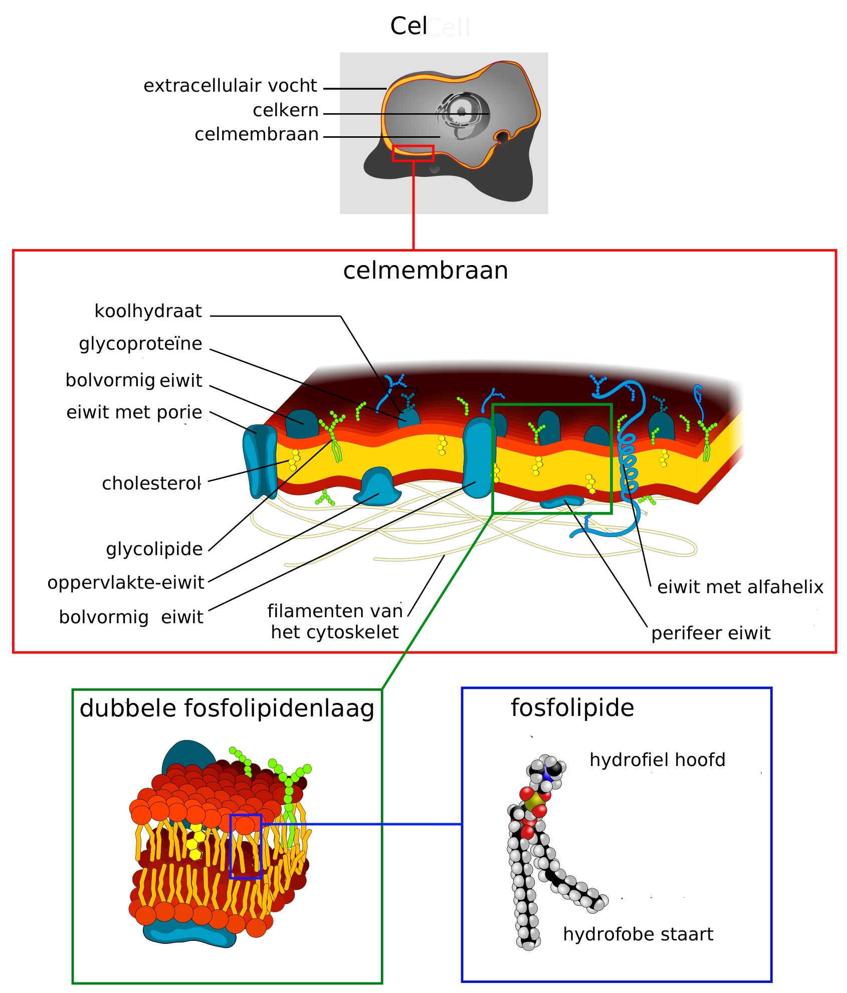 The cell membrane, also called the plasma membrane...