SHC 4113 Human Anatomy
Lecture 1 Anatomical terminologies
Anatomy definition: study of the structure of living organisms: part of the body, form, position and relationship among structures
Anatomical position
Standing erect (upright position)
Eyes are level, directed forwards
Arms at the side with the palms facing forward
Legs are parallel with feet on the floor
Directional terms: Anterior (ventral), Posterior (dorsal), superior (cranial or cephalad), inferior (caudal), medial, lateral, proximal, distal, superficial (external), deep (internal), parietal, visceral, ipsilateral, contralateral
Body planes and sections: sagittal section, frontal (coronal) section, transverse (cross) section
Body movement: flexion (bending), extension, abduction, adduction, circumduction, rotation, elevation, depression, protraction, retraction, pronation, supination
Special movement of the body
Head and trunk: lateral flexion, rotation, mandible (lateral excursion, medial excursion)
Hand and digits: ulnar flexion, radial flexion, radial abduction, palmar abduction of thumb, opposition of thumb, reposition
Foot: dorsiflexion, plantar flexion (inversion, eversion)
Bone marking
Muscle and ligaments: Tuberosity, crest, trochanter, line, tubercle, epicondyle, spine, process
Joints: head, facet, condyle, ramus
Depression and opening
For passage of blood vessels and nerves: groove, fissure, foramen, notch
Others: meatus, sinus, fossa
Lecture 2 Bone: an overview
The skeletal system is the framework of bone and cartilage that protects our organs and allows us to move
Bone is the hardest tissue of the body composed of:
Water: 20% organic matter: 30-40%, give toughness
inorganic matter: 40-50%, calcium phosphate and carbonate, give hardness
Functions of the skeleton
Support: form internal framework, support body, cradles soft organs, give size and shape
Protection: protect soft organs, vital organs, e.g.
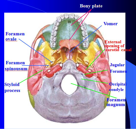 Figure 1
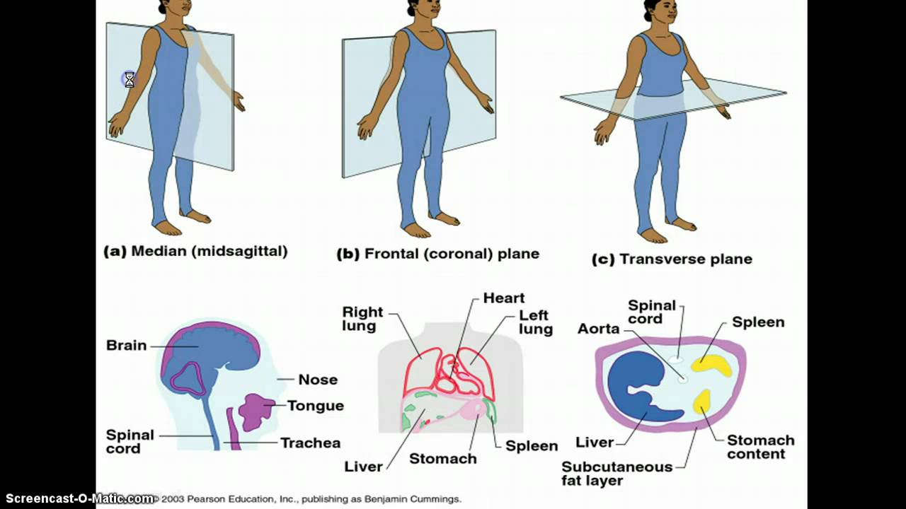 Figure 2
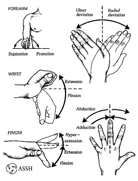 Figure 3
Cranium bones protect the brain
Movement: skeletal muscles attached to bones by tendons, to move the body
Storage: provide storage of mineral salts, e.g. Calcium, phosphorus
Blood cell formation: red bone marrow in cancellous bone makes red blood cells, platelets
Macroscopic structure of bone
Diaphysis: compact bone, give the length of the bone
Periosteum: cover and protect the diaphysis by a fibrous connective tissue membrane
Epiphyses: end of long bone, thin layer of compact bone with an area filled with spongy bone
Articular cartilage: cover the external surface of the bone, smooth, slippery surface to decrease the friction
Epiphyseal line: thin line of bony tissue, remnant of the epiphyseal plate (initial bone development site)
Yellow marrow/ medullary cavity: center of the long bone, high fat content, yellow in color
Medullary canal: hollow space within the diaphysis, contains yellow marrow
Red marrow: fill in the spaces in cancellous bone, rich blood supply, formation of platelets and blood cells
Type of bone tissues (adult skeleton--206 bones)
Compact bone: dense outer layer, solid, hard, ivory in color, covered with fibrous tissue called periosteum
Cancellous bone: found in the extremities of long bones & the interior of small bones, spongy, red marrow filled
Microscopic structure of bone tissue
Compact bone tissue: circular pattern structure, found in the diaphysis of long bone
Haversian canal--containing blood vessels, lymphatics & nerve
Lamellae--concentric plates of bone surrounding the Haversian canal
Lacunae--filled in the spaces of lamellae containing lymph and bone cell
Canaliculi--bring nutrients and oxygen to bone cell and remove waste products
Cancellous (spongy) bone
--red bone marrow is present, red bone marrow is a type of tissue in which blood cells are found & matured before entering the blood vessels
-found in extremities of the long bone (epiphysis) and the inner mass of flat bone
Bone formation (ossification, osteogenesis)
As the cartilage enlarges through appositional and interstitial growth, chondrocytes near the centre of the shaft increase greatly in size. The matrix is reduced to a series of small struts that soon begin to calcify the enlarged chondrocyte then die and disintegrate, leaving cavities within the cartilage.
Blood vessels grow around the edges of the cartilage and the cells of the perichondrium convert to osteoblasts. The shaft of the cartilage then becomes ensheathed in a superficial layer of bone.
Blood vessels penetrate the cartilage and invade the central region. Fibroblasts migrating with the blood vessels differentiate into osteoblasts and begin producing spongy bone at a primary center of ossification. Bone formation then spreads along the shaft toward both ends.
Remodeling occurs as growth continues, creating a marrow cavity. The bone of the shaft becomes thicker, and the cartilage near each epiphysis is replaced by shafts of bone. Further growth involves increase in length.
Capillaries and osteoblasts migrate into the epiphyses, creating secondary ossification centers.
The epiphyses are filled with spongy bone. An articular cartilage remains exposed to the joint cavity; over time it will be reduced to a thin superficial layer. At each metaphysis, an epiphyseal cartilage separates the epiphysis from the diaphysis.
Bone remodeling-old bone is constantly destroyed by osteoclasts, while new bone is formed by osteoblasts
Factors affecting bone growth
Genetic factors-size and shape of bone
Mechanical factors-osteoblast activity increases by muscle contraction(bone tissue thicken, strengthen; osteoclast become active by not enough exercise( bone tissue thinner, weaker
Nutrition-vitamin A necessary for osteoblast, osteoclasts to function; -vitamin C for collagen synthesis and matrix mineralization by osteoblasts, deficiency(growth retard, osteoporosis; -vitamin D for absorbing calcium from intestine, deficiency( rickets, osteomalacia
Hormones --growth hormone: from pituitary gland, increase protein synthesis, promote cell division; increase(gigantism; decrease(dwarfism --thyroid hormone: from thyroid gland, increase rate of replacement of cartilage bon in the growth plate --sex hormone: oestrogen, testosterone stimulate bone growth, and ossification of epiphyseal plate --parathyroid hormone: from parathyroid gland, stimulate osteoclast to breakdown bone, and increased blood calcium levels --calcitonin: from thyroid gland, stop effects of PTH, stop osteoclasts, help calcium absorption by bones
Lecture 3: bones: classification and position
Classification of bones
Long bone
Short bone (cube shape)
Flat bone
Irregular bone
Sesamoid bone (small bone developed from tendon)
Skeleton divided into 2 parts: axial skeleton, appendicular skeleton
Whole body 206 bones; 80 from axial skeleton, 126 from appendicular skeleton
Skull bones-cranium (8), face(14)
Frontal bone, parietal bones, temporal bones (styloid process, mastoid process, jugular foremen), occipital bone, sphenoid bone, ethmoid bone
Foramen magnum allows spinal cord to connect with the brain
Facial bones:14 irregular bones, 12 are paired, only mandible and vomer are single
Maxillae (maxillary bone)
Zygomatic bones (cheekbones)
Lacrimal bones (tears)
Nasal bones
Vertebral column
Structure of intervertebral discs
Cervical vertebrae (7) (C1-C7) atlas and axis are different because they perform functions not shared by the other cervical vertebrae Atlas (C1)-no vertebral body, nod "yes" Axis (C2)-act as a pivot for rotation of the atlas joint between atlas and axis, nod "no" C3- C7 : have small bodies with articular facets for articulation with one another All (C1-C7) have transverse foramina in their transverse processes for the passage of vertebral blood vessels
Thoracic vertebrae (12)-all typical, larger than cervical vertebrae, form a joint with the ribs
Lumbar vertebrae-largest and strongest vertebrae
Sacrum (5)-winglike shape bone
Coccyx (3-5)-tiny, irregular shaped, human "tailbone"
Thoracic cage(bony thorax)-made up of sternum, ribs, thoracic vertebrae, costal cartilages --protective, cone-shaped cage for the organs inside the cavity (heart, lungs, major blood vessels)
Sternum-flat, consists of 3 bones (manubrium, body, xiphoid process)
Ribs-12 pairs true ribs-first 7 pairs, connect to sternum directly by costal cartilages false ribs-next 5 pairs floating ribs--last 2 pairs of ribs, no sternal attachment
Appendicular skeleton
Shoulder girdle (pectoral girdle), consists of 2 bones: clavicle, scapula
--Scapulae (shoulder blades): triangular in shape
Bones of the upper limbs
Arm-humerus
Forearm-radius and ulna
Hand-carpals (8), metacarpals(5), phalanges(14)
Bones of the pelvic girdle
Pelvic girdle: bear the weight of upper body; protect vital organs e.g. reproductive organs, urinary bladder, intestine
Hip bone: ilium, ischium, pubis
Male | Female | |
Inlet | Smaller | Larger, circular |
Pelvis | Heavier, fatter | Shallower, lighter, thinner |
Sacrum | Longer | Shorter |
Ischial spines | Longer | Shorter |
Outlet | Smaller | Larger |
Pubic arch | Not rounded | Rounded |
Angle of pubic arch | Smaller | Greater |
Bones of the lower limbs
Lecture 4: Different types of joints
Joints classification
26 irregular bones, called vertebrae
Spinal nerve pass
33 vertebrae in 2 groups:
True vertebrae separated by intervertebral discs�cervical (7), thoracic (12), lumbar vertebrae (5)
False vertebrae are fixed to one another
Sacrum: 5 sacral vertebrae�coccyx: 4 fused coccygeal vertebrae
�
�
Fixed joints/ fibrous joints
Held by fibrous tissue
Cartilaginous joints/ slightly movable joint
A pad of white fibrocartilage between the ends of bones making up the joint
Freely moveable joints/ synovial joint
Presence of synovial membrane
Synovial joints-with synovial fluid
Structure of a synovial joint
Capsular ligament/ joint capsule
Articular cartilage
Synovial membrane
Extra-capsular structure
Intra-capsular structure