The kidney is made up of nephrons, which are a kidney's functional units. These nephrons collect fluid filtered from the blood. The kidney connects to the renal artery, renal, vein, and ureter. Purified blood leaves the kidney using the renal vein, urine leaves using a ureter and the renal artery carries blood from the aorta to the kidney.
The nephron has a cup-shaped nephric capsule that surrounds a cluster of capillaries called the glomerulus. A good deal of fluid from the blood filters into the capsule. Large proteins and whole blood cells are left behind due to the fact that their too big to pass through the filters along with the plasma or blood fluid.
There are four main parts of the nephron tubule: the proximal convoluted tubule, the U-shaped loop of Henle, the distal convoluted tubule, and the collecting duct. A substantial amount of resorption takes place in the proximal convoluted tubule.
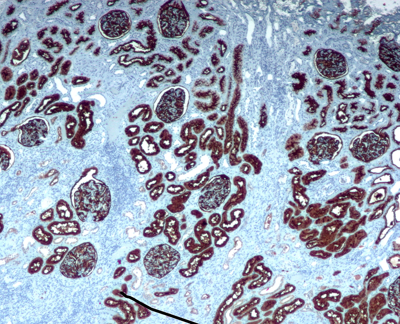 English: Immunohistochemical staining of a non-neo...
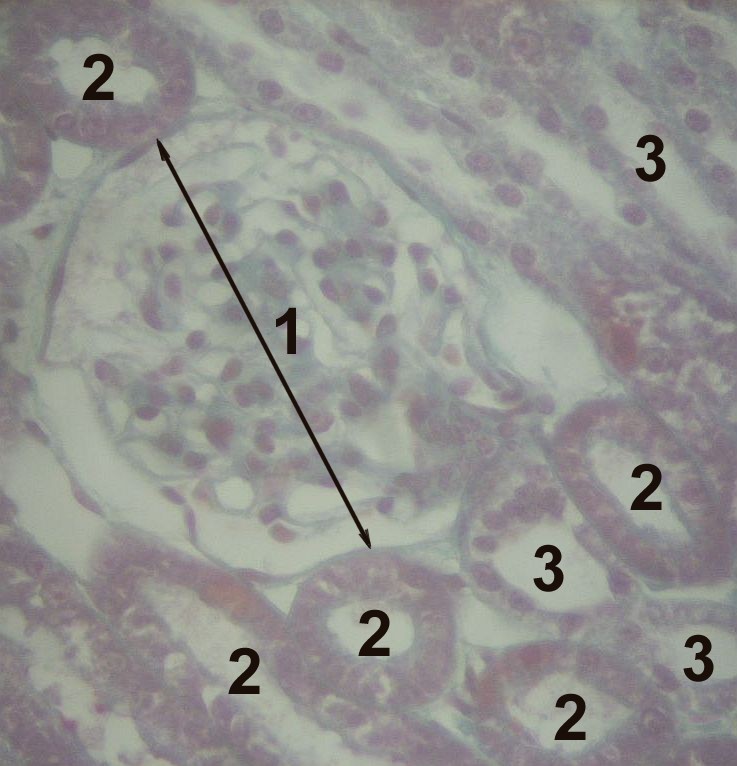 histology of the cortex of a kidney; 1—Glome...
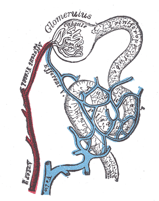 Distribution of blood vessels in cortex of kidney....
The small proteins, glucose, and ions are returned to the blood by active transport. If the glucose in the filtrate, or filtered fluid, exceeds the kidney threshold level, some glucose will remain and appear in the urine. The loops of Henle permit the production of rather concentrated urine. The collecting duct, together with the loop of the Henle, plays a vital role in water balance.
The concentration of urine takes place in the collecting ducts although the process depends on the activities in the loops of Henle. The loops of Henle and the collecting ducts are in the medulla of the kidney. The other parts of the nephron are outside the medulla in the outer region, the cortex. There's a fluid in the medulla that contains an osmotic gradient in which solutes are steadily more concentrated in the direction away from the cortex. There are two...