Running Head: MRI OF SOFT TISSUESMRI of Soft TissuesTable of Contents1. Introduction â¦â¦â¦â¦â¦â¦â¦â¦â¦â¦â¦â¦â¦â¦â¦â¦â¦â¦â¦â¦â¦â¦.â¦â¦.â¦.....â¦â¦.42. Most Conditions Involve Soft Tissue MRI Scanningâ¦â¦â¦â¦â¦â¦â¦â¦...â¦â¦â¦.â¦.43. Breast MRI â¦.................................................................................................................4a.Indication â¦â¦â¦â¦.â¦â¦.â¦.â¦â¦â¦â¦â¦â¦â¦â¦â¦â¦â¦â¦â¦â¦â¦â¦â¦4b.Technical Considerations with Breast MRI â¦â¦â¦â¦â¦â¦.â¦â¦â¦â¦â¦..5c.Patient Positionâ¦â¦â¦â¦â¦â¦â¦â¦â¦â¦â¦â¦â¦â¦â¦â¦â¦â¦â¦â¦â¦â¦...5d.Imaging Protocols Sequences and Techniquesâ¦â¦â¦â¦â¦â¦â¦â¦â¦â¦...6e.Use of Contrast Agentsâ¦â¦â¦â¦â¦â¦â¦â¦â¦â¦â¦â¦â¦â¦â¦â¦â¦â¦â¦...7f.Limitations in Breast MRIâ¦â¦â¦â¦â¦â¦â¦â¦â¦â¦â¦â¦â¦â¦â¦â¦â¦â¦..84. Knee MRI.......................................................................................................................8a.Indication for Knee MRI â¦â¦â¦â¦...â¦â¦â¦...â¦â¦..â¦â¦â¦â¦â¦â¦â¦â¦â¦..9b.Patient Position â¦â¦â¦â¦â¦â¦â¦â¦â¦...â¦â¦.â¦â¦â¦â¦â¦â¦â¦â¦â¦â¦â¦â¦.9c.Pulse Sequences â¦â¦â¦â¦â¦â¦â¦â¦â¦â¦â¦â¦â¦â¦â¦â¦â¦â¦â¦â¦..â¦â¦.â¦.9d.Knee Structures Appearance .â¦â¦â¦â¦â¦â¦â¦â¦â¦â¦â¦â¦â¦.â¦â¦â¦â¦â¦...9e.Knee Ligaments â¦â¦...â¦â¦â¦â¦.â¦â¦â¦â¦â¦â¦â¦â¦â¦â¦â¦â¦â¦â¦â¦â¦...10Anterior Cruciate Ligament (ACL)â¦â¦â¦â¦â¦.â¦â¦â¦â¦â¦â¦â¦â¦...10Posterior Cruciate Ligament (PCL)â¦â¦â¦â¦â¦â¦â¦â¦â¦â¦â¦â¦â¦â¦105. Shoulder MRI â¦â¦â¦â¦â¦â¦â¦.â¦.â¦â¦â¦â¦â¦â¦â¦â¦â¦â¦â¦â¦..â¦â¦â¦.â¦â¦â¦â¦11a.Indications for Shoulder MRIâ¦â¦â¦.â¦â¦â¦â¦â¦â¦â¦..â¦â¦.â¦â¦................11b.Patient Position â¦â¦â¦â¦â¦â¦.â¦â¦â¦â¦â¦â¦â¦â¦â¦â¦â¦â¦â¦â¦.................11c.Area Problems â¦â¦â¦â¦â¦â¦â¦â¦â¦â¦.â¦â¦â¦â¦.â¦â¦...â¦â¦â¦â¦â¦â¦â¦..11d.Imaging Protocols Sequences and Techniquesâ¦â¦â¦â¦â¦â¦â¦â¦â¦â¦â¦â¦..12e.Normal Shoulder MRI â¦â¦â¦â¦â¦â¦â¦â¦â¦â¦â¦....â¦â¦...............................12f.Abnormal Shoulder MRIâ¦â¦â¦â¦â¦â¦â¦â¦â¦â¦â¦â¦â¦â¦â¦â¦â¦â¦â¦â¦â¦12g.Rotator Cuff MRIâ¦â¦â¦â¦â¦â¦â¦â¦â¦â¦â¦â¦â¦â¦â¦â¦â¦â¦â¦â¦â¦â¦â¦â¦13Differentiation of Rotator Cuff Tendon Pathology..â¦â¦â¦â¦â¦â¦â¦...146. Elbow â¦â¦â¦â¦â¦â¦â¦â¦â¦â¦â¦â¦â¦â¦â¦â¦â¦â¦â¦â¦â¦â¦...â¦â¦â¦â¦â¦â¦â¦â¦â¦.15a. Technique â¦â¦â¦â¦â¦â¦â¦â¦â¦â¦â¦.â¦â¦â¦...................................................15b. Elbow Soft Tissue & Ligaments â¦â¦â¦â¦â¦â¦â¦â¦â¦â¦â¦â¦â¦â¦â¦....â¦â¦..167. Masses in different location in the body â¦â¦â¦â¦â¦â¦â¦â¦â¦â¦â¦â¦â¦â¦â¦..â¦â¦â¦..17a. Thigh MRIâ¦â¦â¦â¦â¦â¦â¦â¦â¦â¦â¦â¦â¦â¦â¦â¦â¦â¦â¦â¦â¦â¦â¦â¦â¦â¦â¦.17Patient Positionâ¦â¦â¦â¦â¦â¦â¦â¦â¦â¦â¦â¦â¦â¦â¦â¦â¦â¦â¦â¦â¦â¦17Pulse Sequencesâ¦â¦â¦â¦â¦â¦â¦â¦â¦â¦â¦â¦â¦â¦â¦â¦â¦â¦â¦â¦â¦...17b. Legâ¦â¦â¦â¦â¦â¦â¦â¦â¦â¦â¦â¦â¦â¦â¦â¦â¦â¦â¦â¦â¦â¦â¦â¦â¦â¦â¦â¦â¦....19Patient Positionâ¦â¦â¦â¦â¦â¦â¦â¦â¦â¦â¦â¦â¦â¦â¦â¦â¦â¦â¦â¦â¦â¦.19Pulse Sequences and Techniqueâ¦â¦â¦â¦â¦â¦â¦â¦â¦â¦â¦â¦â¦â¦â¦â¦19Focal Myositis in the Legâ¦â¦â¦â¦â¦â¦â¦â¦â¦â¦â¦â¦â¦â¦â¦â¦â¦â¦..198. Case study â¦â¦â¦â¦â¦â¦â¦â¦â¦â¦â¦â¦â¦â¦â¦â¦â¦â¦â¦â¦....â¦â¦â¦â¦â¦â¦â¦â¦â¦...20a. Case study 1 (MRI Left-Knee)â¦â¦â¦â¦â¦â¦â¦â¦â¦â¦â¦â¦â¦â¦â¦â¦â¦â¦â¦â¦â¦21Introduction â¦â¦â¦â¦â¦â¦â¦â¦â¦â¦â¦â¦â¦â¦â¦â¦â¦â¦â¦â¦â¦â¦.â¦..21Clinical History Detailsâ¦â¦â¦â¦â¦â¦â¦â¦â¦â¦â¦â¦â¦â¦â¦â¦â¦â¦â¦..21Preparation â¦â¦â¦â¦â¦â¦â¦â¦â¦â¦â¦â¦â¦â¦â¦â¦â¦â¦â¦â¦â¦.â¦â¦â¦21Technique â¦â¦â¦â¦â¦â¦â¦â¦â¦â¦â¦â¦â¦â¦â¦â¦â¦â¦â¦â¦â¦.â¦..â¦â¦21Protocol
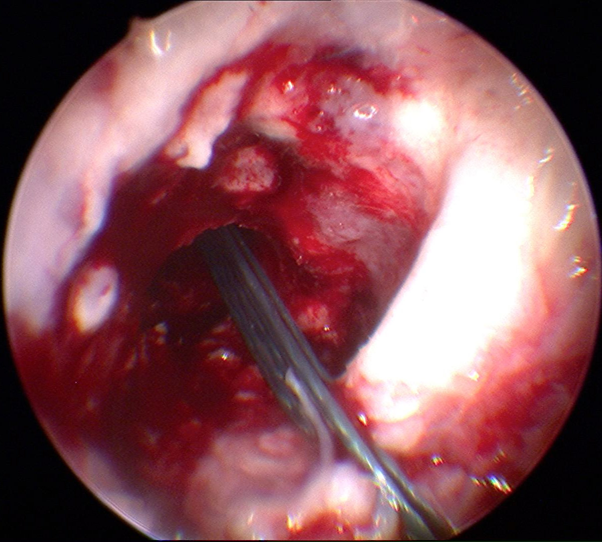 English: Arthroscopic reconstruction of the anteri...
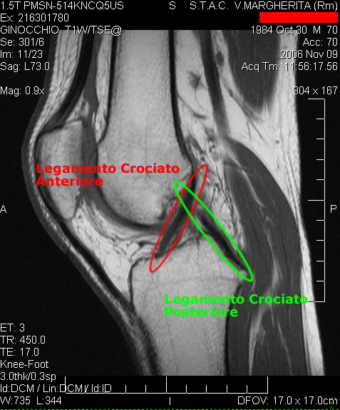 Cruciate ligaments
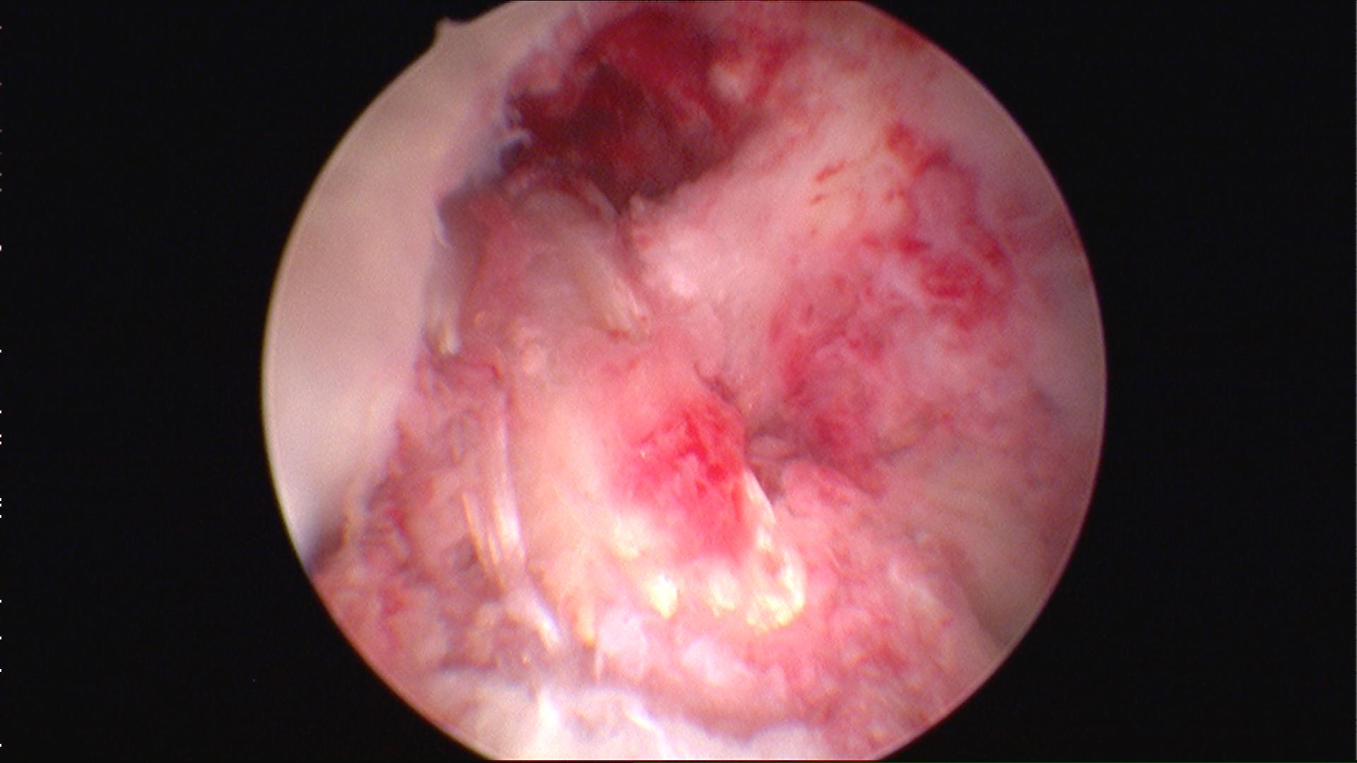 Arthroscopic reconstruction of the anterior crucia...
â¦â¦â¦â¦â¦â¦â¦â¦â¦â¦.â¦..â¦â¦â¦â¦â¦â¦â¦â¦â¦â¦â¦â¦â¦â¦21Findingsâ¦â¦â¦â¦â¦â¦â¦â¦â¦â¦â¦â¦â¦â¦â¦â¦â¦â¦â¦â¦â¦â¦â¦â¦â¦â¦23Commentâ¦â¦â¦â¦â¦â¦â¦â¦â¦â¦â¦â¦â¦â¦â¦â¦â¦â¦â¦â¦â¦â¦â¦â¦â¦..23b. Case study 2 (MRI Shoulder - Right)â¦â¦â¦â¦â¦â¦â¦â¦â¦â¦â¦â¦â¦â¦â¦..â¦â¦â¦..24Introduction â¦â¦â¦â¦â¦â¦â¦â¦â¦â¦â¦â¦â¦â¦â¦â¦â¦..â¦â¦â¦..â¦â¦â¦.24Clinical History Detailsâ¦â¦â¦â¦â¦â¦â¦â¦â¦â¦â¦â¦â¦â¦â¦â¦â¦â¦â¦â¦24Preparation â¦â¦â¦â¦â¦â¦â¦â¦â¦â¦â¦â¦â¦â¦â¦â¦â¦â¦â¦â¦â¦..â¦..â¦..24Techniqueâ¦â¦â¦â¦â¦â¦â¦â¦â¦â¦â¦â¦â¦â¦.â¦â¦â¦â¦â¦â¦â¦â¦â¦â¦â¦24Protocolâ¦â¦â¦â¦â¦â¦â¦â¦â¦â¦â¦â¦â¦â¦â¦â¦â¦â¦â¦â¦â¦â¦â¦â¦â¦â¦.25Findingsâ¦â¦â¦â¦â¦â¦â¦â¦â¦â¦â¦â¦â¦â¦â¦â¦â¦â¦â¦â¦â¦â¦â¦â¦â¦â¦26Commentâ¦â¦â¦â¦â¦â¦â¦â¦â¦â¦â¦â¦â¦â¦â¦â¦â¦â¦â¦â¦â¦â¦â¦â¦â¦..269. Conclusion â¦â¦â¦â¦â¦â¦â¦â¦â¦â¦â¦â¦â¦â¦â¦â¦â¦â¦â¦â¦â¦â¦â¦..â¦.â¦â¦â¦â¦â¦â¦..2710. References â¦â¦â¦â¦â¦â¦â¦â¦â¦â¦â¦â¦â¦â¦â¦â¦â¦â¦â¦â¦â¦â¦â¦â¦â¦â¦â¦â¦â¦â¦â¦28IntroductionWith the assistance of advanced hardware and software, Magnetic Resonance Imaging (MRI) modality generates effective magnetic and radio waves, which are used by the radiologists for producing elaborated images of the inner parts of the body. MRI modality has improved the technology of radiology by not exposing the patients and personnel to the high frequency radiations harmful for the body. Along with this, the capability of diagnosing a patient through MRI has improved the quality and resolution of the images, which effectively and accurately distinguishes the normal and abnormal soft tissues of the body (Breitenseher, 2004).
MR images provide better images of the soft tissues than the other imaging modalities. In the assessment of the soft tissues and masses of joints, MRI modality is effective. For generating more clear and accurate images, contrast agents are...
MRI of Soft Tissues
Well structured paper.
0 out of 0 people found this comment useful.