1. Describe the anatomy and function of the human eye.
Conjunctiva- a delicate membrane that covers the surface of the
eye and the inside of eyelids.
Protects the front of the eye.
Cornea- The front part of the eyeball. It is transparent, relatively
thick. Bends light rays as they pass through it.
Sclera- Continuous with the cornea but not transparent. Forms
tough white outer back part of eyeball. Protects the eye, helps maintain
it's shape.
Choroid- lies on the inside of sclera, think black pigmented layer
containing blood vessels. Pigment absorbs stray light preventing false
images.
Retina- Innermost layer of eye. Lines back of eyeball, contains
light sensitive cells or photoreceptors and nerve fibres. Receives the light
and changes it in to electrical impulses that travel to the brain via the optic
nerve.
Iris- coloured part at front of eye, composed of muscles.
Regulates the amount of light entering the eye.
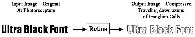 English: A graphical depiction of the edge detecti...
 A photo showing refraction of light rays: a soda s...
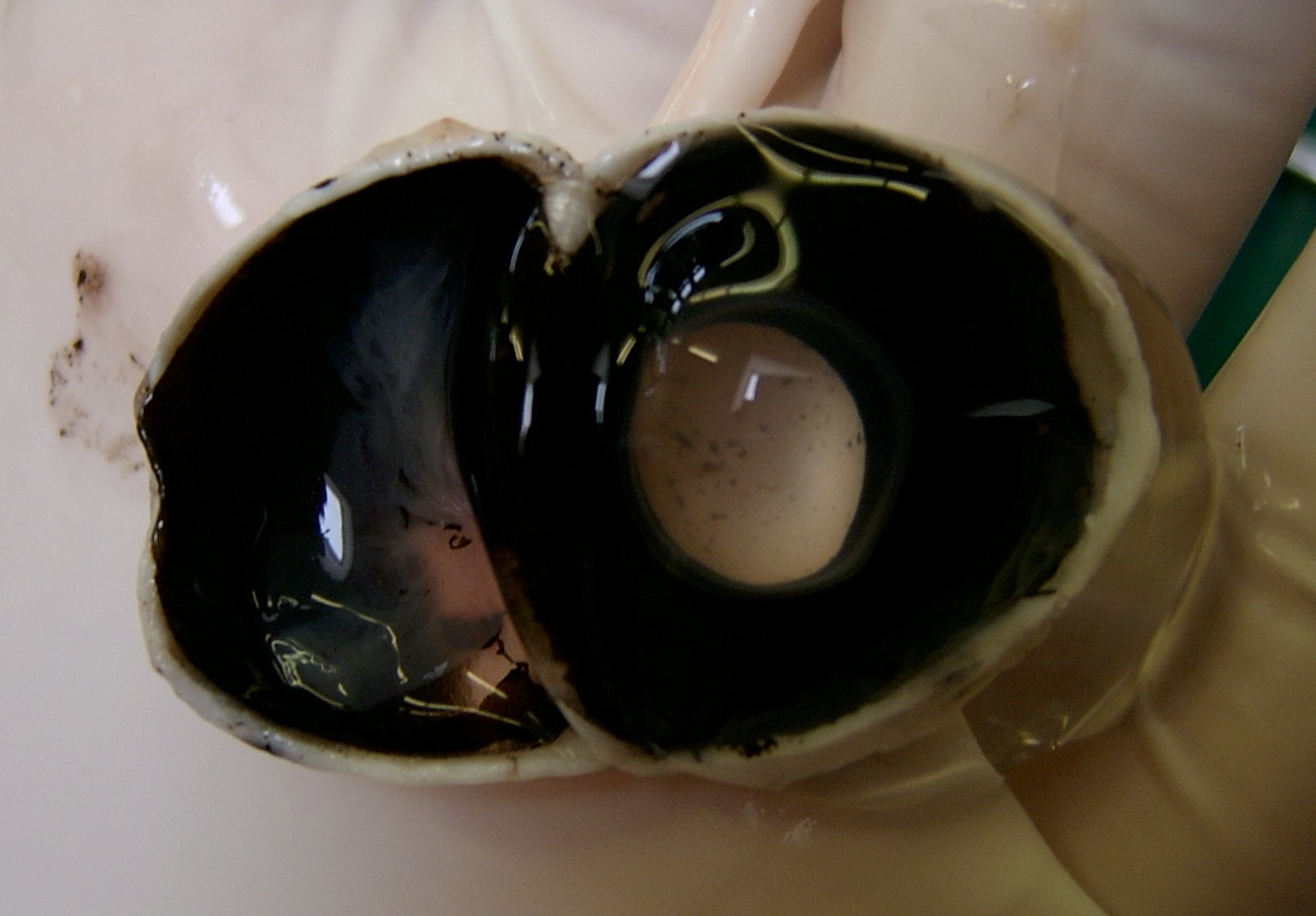 You see the cut open eyeball. Note the "zooming" e...
Lens- transparent biconvex protein disc behind the pupil. Focuses
light rays on to the retina.
Aqueous humor and Vitreous humor- aqueous humor is viscose
liquid that fills the front chamber of the eye. Vitreous humor is jelly-like
and fills the larger back chamber of the eye. Help keep the eyeball in
shape and also bend rays of light as they pass through.
Ciliary body- connects the choroid with the lens. It contains
suspensory ligaments and ciliary muscles. Ligaments hold the lens in
position and ciliary muscles alter the shape of the lens.
Optic nerve- connects eye with brain. The region where the optic
nerve leaves the eye is known as the blind spot because it has no
photoreceptors so it can't produce an image. Carries nervous signals from
retina to visual cortex of the brain, which interprets them as, images.
2. a) Explain the term 'refraction' in...