Dogfish Shark Dissection
You will complete this guide and turn it in for your project grade
I. Predissection notes
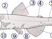
Label the following six anatomical positions: on the diagrams of the sharks below.
Cranial -
Caudal -
Dorsal -
Ventral-
Medial-
Distal-
Science 17 January 2003: Vol. 299 no. 5605 pp. 389-392 DOI: 10.1126/science.1079777
Collapse and Conservation of Shark Populations in the Northwest Atlantic
A. External Anatomy
Use your guide to define the following parts of the external anatomy of your dogfish.
Rostrum- Where is it found?________________________________________________
_______________________________________________________________________
What does it help the shark to do? ___________________________________________
Nares- What are these similar to in humans? ___________________________________
Where are they located? ___________________________________________________
When water travels up the nares it reaches an organ that allows it to achieve a legendary feat.
What is the part of the body that it reaches and what does this allow the shark to do?
Jaws- How many pounds of pressure per sq inch can a shark exert?
What is interesting about the sharks teeth and scales?
Eyes- What is the name of the outer covering of the eyes?___________
What does the contraction of the iris adjust?
Spiracles- How do these openings allow the shark to stay alive?
Gill Slits- Remember these are what forms from the pharyngeal slits present during a sharks development. Note that there is no outer covering or Operculum like there are in bony fishes.
Where does water pass through before it passes over the internal gills?
Pectoral,
Pelvic,
and
Cloaca fins
Use the diagrams on pages 12 & 13 to determine if it is a male or female.
Do you have a male or a female?
What do sharks use claspers for?
B. Skeletal System
Sharks have a skeletal system that is made up of cartilage. They do not actually ever develop bone. There are many interesting hypotheses about how bone arose in evolution. One prominent hypothesis is that at some point a mutation took place in the DNA that lead to the way that cartilage developed. This mutation lead to a change in how cartilage interacted with different chemicals in the ocean. One of these chemicals phosphate has a tendency to harden when it is stored in cartilage. If this did happen in a ancestor predating sharks and gnathostomes it would have been very advantageous to the survival of those "lucky" mutants, because phosphorous is an essential chemical component for survival.
This hypothesis of a mutation that gave rise to phosphorous bearing cartilage is all the more plausible in light of several mutations that take place in humans. For example, Primrose Syndrome is a rare genetic disease where individuals have a side effect that their ear cartilage becomes laden with phosphorous and turns into bone. This can also happen in Fibrodysplasia ossificans progressiva or stone- man syndrome. Again, one of the side effects of this is that a mutation leads to the body's building of bone in place of cartilage tissue. Amazingly, a chemical compound Squalamine taken from dogfish is sometimes used to treat the bone build up in patents who suffer from Fibrodysplasia ossificans progressiva!
C. Muscular System
What is the Origen of a muscle-
What is the insertion of a muscle?
-So, if a shark has a muscle that moves it's pectoral fin up or down the origen is where this muscle is anchored in the shark's body and the insertion is where it attaches to the pectoral girdle in order to move it up or down.
It is really a fun experience to dissect down to a single muscle and watch what it moves when it is pulled or relaxed.
How are your triceps and biceps an example of antagonistic pair?
Define Flexion-
Define Extension-
Flex and extend the pectoral fin
Myotomes are arranged in a curious pattern. What is that pattern?
What separates one myotome from another?
What does a septum do?
On your diagram of anterior muscles indicate where the following parts are.
Epaxial myotome - hypaxial myotome - Adductor Mandibulae - Spiracularis-
What are the superficial constrictors primarily involved in?
What do the levatator muscles primarily help with?
Can you find one?
What do the interarcuals primarily act upon?
Appendicular Muscles
Fins
Locate the flexor and extensor and label it on your diagram
Locate your pectoral flexor and extensor on your shark- demonstrate flexion and
extension to get Mr. Smalley's initials _______
IV. Dissecting the Abdominal Cavity
For this section follow the lab guide to help you open your shark. You should locate the major organs
heart
liver
stomach
spleen
intestine
duodenum
spiral intestine
colon
pancreas
gall bladder
kidney
brain
gonads female: ovaries male: testes
Sketch the Internal Anatomy of the Dogfish Shark (10 points)
Sketch the internal anatomy of the dogfish shark in the space below. Use the lab manuals to help you label at least 10 recognizable structures in your sketch.
Post-Lab - Answer the following questions before continuing with your dissection:
1. How is the shark's digestive system different from a human's?
2. How is the shark's circulatory system different from a human's?
3. How did you position your shark to begin dissecting the abdominal cavity?
4. Why do you think cutting through the pectoral girdle may have been difficult?
5. Write at least one think you found interesting about the shark's digestive or circulatory system.
There will be a lab practical where you will be expected to be able to locate the following structures: (10 structures will be drawn at random for each student.)
Head
nares (nostrils), mouth, eyes, spiracles, external gill slits, ampullae of Lorenzini, lateral line system
Trunk
dorsal fins, pectoral fins, pelvic fins, cloaca, anus, male: claspers, caudal fin
Major Organs
heart, liver, stomach, spleen, intestine, duodenum, spiral intestine, colon, pancreas, gall bladder, kidney, gonads, brain, female: ovaries, male: testes
Major Muscles
Adductor Mandibulae, Epaxial myotome, hypaxial myotome, hynoid constrictors, pectoral levator


