INTRODUCTIONTeratogens are chemicals, infectious agents, physical conditions, or deficiencies that, on fetal exposure, can alter fetal morphology or subsequent function (Chung, 2005). The effect of a teratogen on the developing organism depends on what period in development the embryo is exposed to the teratogen (Jaeckel, 2001). The effect of a teratogen on the developing organism also depends on the dose and/or frequency of exposure of/to the teratogen (Jaeckel, 2001). Another factor that determines whether a specific teratogen will be harmful is the genetic make-up of the developing organism (Jaeckel, 2001). Possessing or lacking certain genes makes the developing organism more susceptible to the effect of a teratogen.
The purpose of this experiment is to examine the effects of teratogens, whether deleterious chemicals or physical environmental factors, on embryonic development of Xenopus Laevis. Xenopus Laevis is a South African clawed frog, containing many features that make it widely used as a model organism in developmental biology.
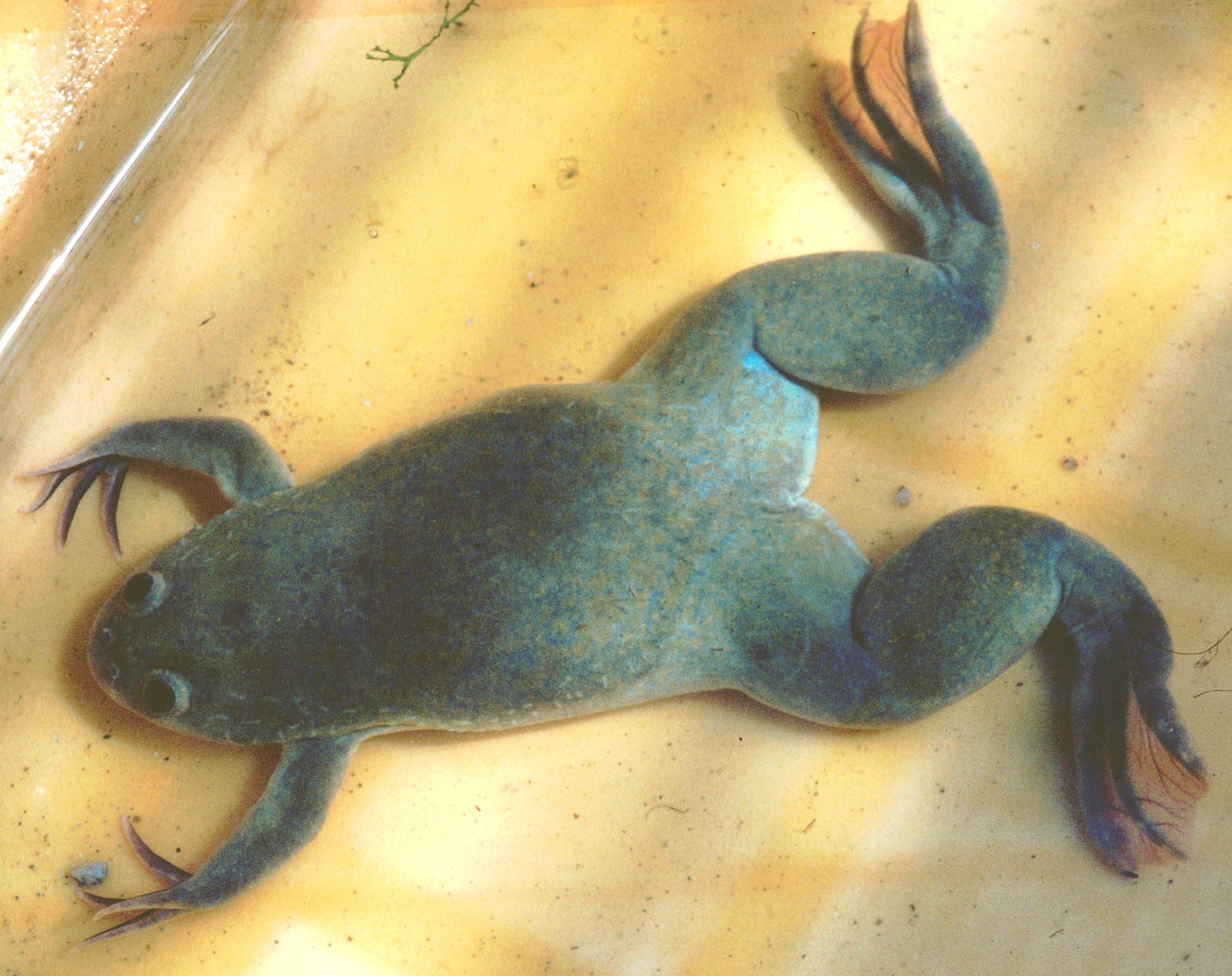 Xenopus laevis
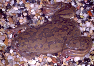 Xenopus
 Lafe Gleason & wife & J. Jaeckel (LOC)
In Xenopus embryos, the maternal contribution to early embryogenesis is considerable (Kay & Peng, 1991). The fertilized egg develops to the late blastula stage before significant transcription of the embryonic genome occurs (Kay & Peng, 1991). It follows, therefore, that cytoplasmic stores of proteins and messenger RNAs accumulated during oogenesis are necessary after fertilization for development of the embryo to the late blastula stage.
The four compounds used as teratogens in this experiment are retinoic acid, lithium chloride, caffeine, and ethanol. Retinoic acid is known to produce a concentration-dependent series of defects in anterior axial structures that range from small deletions to embryo lacking heads (Altaba & Jessell, 1991). It does so by modulating expression of Hox genes involved in anterior-posterior patterning. Lithium chloride is shown to induce dorsalization in early-stage embryos (Spenillo, 2001). Lithium chloride has the ability to inhibit glycogen synthase kinase-3, which is a substance that initiates the wnt-pathway leading to dorsal axis formation (Spenillo, 2001). Lithium chloride also inhibits enzymes participating in the hydrolysis of intermediate inositol phosphates, inositol monosphosphate phosphatase, and inositol polyphosphate 1-phosphatase, thus blocking the recycling of IP-3 into inositol (Kume et al., 2000).
Caffeine interferes with the development of Xenopus larvae in a concentration-dependent manner. It induces characteristic external abnormalities, such as shortened body and wavy fins (SAKAMOTO, 1993). It was revealed that exposure to caffeine induced severe damage in the myotome and neural tube, and at higher concentrations, the epidermal tissue was also affected (SAKAMOTO, 1993). Finally, ethanol induces a behavioral dysfunction, such as the fetal-alcohol syndrome (Lindi et al., 2001).
PROCEDURE(Obtained from lab protocol prepared by Dr. Plenefisch)This lab has a total of seven groups. Thus, two groups studied the effects of lithium chloride, two groups studied the effects of caffeine, two groups studied the effects of ethanol, and one group, which was our group, studied the effects of retinoic acid. Four exposure conditions were picked, plus a control (untreated) condition. Thus, five plates of embryos were prepared. Four of them were treated with a compound, and one was untreated. Each plate contained four embryos. Embryos were separated from the egg mass and washed in sterile pond water. Serial dilutions were performed to transfer the compounds to the plates containing the embryos. Those are shown in Figure (1) below. The solvent in all of the tubes, except the retinoic acid tube, was pond water. The solvent in the retinoic acid tube was methanol instead of pond water. Plate #5 was a control plate, and thus, only contained 20mL of pond water.
Figure (1): Serial Dilutions of TeratogensAll of the plates tested for time and concentration except the retinoic acid plate, which only tested for concentration. After several days, the experimental and control Embryos were examined for Viability, size, head size, neutral Tube closure, eyes, suckers, and spinal curvature. Any other noticeable differences or defects between the groups of embryos were noted.
RESULTSAs just mentioned, the effects of retinoic acid, lithium chloride, caffeine, and ethanol on Xenopus development were tested by exposing these compounds at different times as well as in different concentrations. Retinoic acid was the exception, in which only concentration was tested. After careful examinations of the experimental and control embryos, results were obtained and are presented in the tables below. Concentration treatments are presented in Table (1) and time treatments are presented in Table (2).
Table (1): Variations of Concentrations of Teratogens Have Different Effects on Xenopus DevelopmentUndiluted0.47% of compoundSerial Dilution #10.047% of compoundSerial Dilution #20.0047% of compoundSerial Dilution #30.00047% of compoundControlRetinoic Acid0/4 developed. Cell migration halted0/4 developed. All mold with signs of morphogenesis showing0/4 developed. No eggs were transferred to this plate0/4 developed. 1 molded egg and 3 eggs showed developed tadpoles inside1/4 developed. 3 molded eggs and one 1 developed lacking proper locomotion.
Lithium Chloride4/4 growing1/4, development halted after 6 days0/4, development halted after 5 daysN/A3/4 developedCaffeine4/4 living tadpoles3/4 living and 1 dead egg with mold1/4 living, 1 dead, and 2 molded eggs2/4 living tadpoles, 1 dead, and 1 molded egg1/4 living, 2 dead, and 1 molded egg.
Ethanol3/4 developed, but slow reacting4/4 developed, but slow reacting4/4 developed, and function normallyN/A4/4 developed, and function normallyTable (2): Variations of Exposure Times of Teratogens Have Different Effects on Xenopus Development3 Hours1 Day2 Days5 Days7 DaysLithium ChlorideN/ADeveloped like the control but no pigmentation observedNo outer membrane in LiCl eggs. Development was stiff and no curvature like the control. All were dead. No formation of tadpole was observed. Complete disassociation of the membranesN/ACaffeineAll dead. No development3/4 alive, smaller heads and darker neural tubeN/A2/4 alive, smaller and skinnier heads/bodiesN/AEthanolN/A16/16, normal development compared to control3/4 of control eggs developed. 9/16 of ethanol eggs developed. 1/4 of control eggs developed. 8/16 of ethanol eggs developedSame as ÃÂ5 daysÃÂ but more developedDISCUSSIONAs previously stated, the purpose of this experiment was to examine the effects of teratogens, whether deleterious chemicals or physical environmental factors, on embryonic development of Xenopus Laevis. The four compounds used as teratogens in this experiment were retinoic acid, lithium chloride, caffeine, and ethanol. The effects of these compounds were tested at different concentrations (serial dilutions) as well as at different time periods (3 hours, 1 day, 2days, 5days, and 7days).
First, letÃÂs look at the results obtained from the retinoic acid treatment. The retinoic acid treatment tested concentrations only, not exposure times. It can be seen that the more concentrated the retinoic acid, the more harmful it was on Xenopus development. When retinoic acid became more diluted as progressing through the serial dilutions, it became less harmful and some development within the eggs was shown. This explains the results, which showed no development in plates #1-3 but little development within the eggs in plate #4 and a functioning adult tadpole in plate #5 (the control).
On a molecular level, Retinoic acid affects the anterior-posterior patterning of the body by modulating expression of the Hox genes. Thus, modulation of Hox genes could have caused fetal homeotic transformation, leading to death. Also, high concentrations of retinoic acid could have had more effect on Hox genes than low concentrations. This supports the idea mentioned earlier, that the effect of a teratogen on the developing organism does indeed depend on the dose and/or frequency of exposure of/to the teratogen. Thus, the control plate should have had all four developed tadpoles as it contained no retinoic acid. However, the fact that methanol was added to the plates instead of pond water (the usual habitat for Xenopus Laevis), could have caused effects on development as well.
Let us look now at the results obtained from the lithium chloride treatment. The lithium chloride treatment tested both concentrations and exposure times. Table (1) shows that concentrated levels of lithium chloride resulted in all four tadpoles developing. However, as lithium chloride became more diluted, development was restricted to one developed tadpole in the 1st serial dilution plate and no development at all in the 2nd serial dilution plate. These results are inconsistent with the idea that the more concentrated the teratogen, the more harmful it will be. Mold formation on the eggs could have restricted them from developing.
The results in Table (2), however, are more consistent. Table (2) shows that the longer the exposure time to Lithium chloride, the more defects resulted in the embryos such as lack of pigmentation, lack of outer membranes, and even death in cases where exposure time was too long. Thus, this is supported by the fact that after five days of exposure to lithium chloride, no tadpoles developed. The reason lithium chloride was harmful is because it has the ability to inhibit glycogen synthase kinase-3, which initiates the wnt pathway in Xenopus embryos that leads to dorsal axis formation. Thus, lithium-induced embryos underwent dorsalization, altered development, and death as shown by the obtained results.
Caffeine was the third compound to have its effects tested based on concentrations as well as exposure times. Looking at Table (1), the results also do not seem consistent. It looks like the less concentrated the caffeine was, the higher the death rates among the embryos. Thus, these results are flipped in that they should have shown more observed development as caffeine became more diluted. Again, this could be a result of using non-sterile techniques during the experiment, leading to the formation of mold around the cells and restricting development.
Likewise, the results in Table (2) are inconsistent with the idea that longer exposure times should result in more defects and more deaths. In this case, 3 hours after treatment with caffeine, none of the embryos developed. 5 days after exposure, on the other hand, resulted in two developing tadpoles. Again, this could be due to human errors or contamination in the plates that caused restricted development of the frog embryos. Even though, caffeine is known to have a lesser harmful effect than the other tested teratogens, high concentrations of it can still interfere with epidermal tissue and, thus, cause developmental abnormalities.
Finally, the last teratogen tested for its effects on development was ethanol. The results obtained from the ethanol treatment are more consistent in comparison with the caffeine results. Looking at Table (1), we can see by comparing the undiluted plate with the 2nd serial dilution plate that as ethanol became more diluted, more functional tadpoles developed. Likewise, Table (2) shows that the longer the embryos were exposed to ethanol, the higher number of deaths was observed. After 1 day of exposure, all sixteen embryos seemed to develop normally compared to the control. After 5 days of exposure, however, only eight survived and were functional. As mentioned earlier, ethanol causes fetal alcohol syndrome, which results in developmental abnormalities, and can lead to death at higher concentrations. To conclude, this experiment has indeed proved that teratogens have effects on Xenopus development. Moreover, these effects depend on the concentrations of the teratogens as well as the teratogensÃÂ exposure times.
Works CitedChung, Wendy. ÃÂTERATOGENS AND THEIR EFFECTS.ÃÂ Columbia University . N.p., 2005. Web. 25 Oct. 2009. .
Jaeckel, Jennifer. ÃÂTeratogens .ÃÂ University of Michigan. N.p., 28 Mar. 2001. Web. 25 Oct. 2009. .
Jessell, T, and Ruiz I Altaba. ÃÂRetinoic acid modifies mesodermal patterning in early Xenopus embryos.ÃÂ Genes and Development . Cold Spring Harbor Laboratory Press, n.d. Web. 26 Oct. 2009. .
Kay, Brian K, and H Benjamin Peng. Xenopus laevis: practical uses in cell and molecular biology Volume 36 of Methods in cell biology Xenopus Laevis: Practical Uses in Cell and Molecular Biology. N.p.: Academic Press, 1991. N. pag. Google Books. Web. 26 Oct. 2009. .
Lindi, Clara, and Et al. ÃÂEFFECT OF ETHANOL EXPOSURE ON XENOPUS EMBRYO LIPID COMPOSITION.ÃÂ Oxford Journals. N.p., 2001. Web. 26 Oct. 2009. .
Mikoshiba, Katsuhiko, Takeo Saneyoshi, and Shoen Kume. ÃÂDesensitization of IP3-induced Ca2+ release by overexpression of a constitutively active Gqa protein converts ventral to dorsal fate in Xenopus early embryos.ÃÂ InterScience. N.p., n.d. Web. 25 Oct. 2009. .
SAKAMOTO, M. ÃÂTeratology .ÃÂ inist. N.p., 1993. Web. 25 Oct. 2009. .
Spenillo, Justin A. ÃÂDEVELOPMENTAL EFFECTS OF LITHIUM CHLORIDE ON XENOPUS EMBRYOS.ÃÂ Smarthmore. Swarthmore College, 6 Apr. 2001. Web. 26 Oct. 2009. .
 Kay Hernan and Emmett Kelly: Palm Beach, Florida
 Figure 1
 Figure 5