What is an image? An optical appearance; a mental representation an idea or conception. The term image may be applied to a picture such as a photograph a painting or a sketch which has a real physical existence. May also be applied to an idea or concept which has a mental rather than physical existence.
Visual images are of two types; Real Images- those having a real physical existence such as a photographic image. Mental Images; Those generated as a mental picture within our own minds and which are accessible only to subjective study.
Real images consist of patterns of light intensity and possibly variations in colour. The patterns of light are created in one of three ways;
Viewing by reflected light from a surface; Reflected light- ultrasound, RNI, Theatre, thermal imaging paper. (pg7)
Viewing by light transmitted through a semitransparent layer; Radiographs, CT, MRI, DIGITAL, Fluoroscopy, ultrasound.
Viewing by light emitted from a fluorescent layer, Fluoroscopy, ultrasound, CT, RNI, Digital, MRI, anglography, endoscopy.
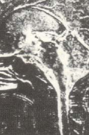 English: Systolic time data acquisition MRI image
 Urban Scene
 Day 294 - West Midlands Police - Film project with...
CONTRAST; To be able to identify a feature or an image, must appear different from its surroundings. On a radiograph, a structure must be of different optical density (Shade of grey) from adjacent structures. If the differences of brightness is great, the structure will stand out well from its surroundings and we say the contrast is HIGH.
HIGH CONTRAST; With a high level of contrast the image produced appears harsh with lots of dense blacks and brilliant whites. Detail may be lost in highlighted areas and in the deep shadows but the boundries between the dark and light areas appear sharply defined.
LOW CONTRAST; With the contrast level at minimum the image appears dull, lacking in any true black and whites. However, inspection of the highlighted areas and the shadows reveal detail that was absent in...