A microscope is an invention that helps us look more closely at a tiny object. Zaccharias Janssen and his father Hans invented the Microscope in 1590. Microscopes are now used in schools, Medical facilities and research facilities and have changed the way we look at things. These are the parts of the microscope Specimen control - hold and manipulate the specimen, stage - where the specimen rests, clips - used to hold the specimen still on the stage micromanipulator device that allows you to move the specimen in controlled, small increments along the x and y axes, Illumination - shed light on the specimen (The simplest illumination system is a mirror that reflects room light up through the specimen.), lamp - produces the light, rheostat - alters the current applied to the lamp to control the intensity of the light produced, condenser - lens system that aligns and focuses the light from the lamp onto the specimen, diaphragms - placed in the light path to alter the amount of light that reaches the condenser Lenses - form the image, objective lens - gathers light from the specimen and magnify the image which is made even larger when we see it through the eyepiece lenses, eyepiece - transmits and magnifies the image from the objective lens to your eye, nosepiece - rotating mount that holds many objective lenses, tube - holds the eyepiece at the proper distance from the objective lens and blocks out stray light, Focus - position the objective lens at the proper distance from the specimen, coarse-focus knob - used to bring the object into the focal plane of the objective lens, fine-focus knob - used to make fine adjustments to focus the image, arm - curved portion that holds all of the optical parts at a...
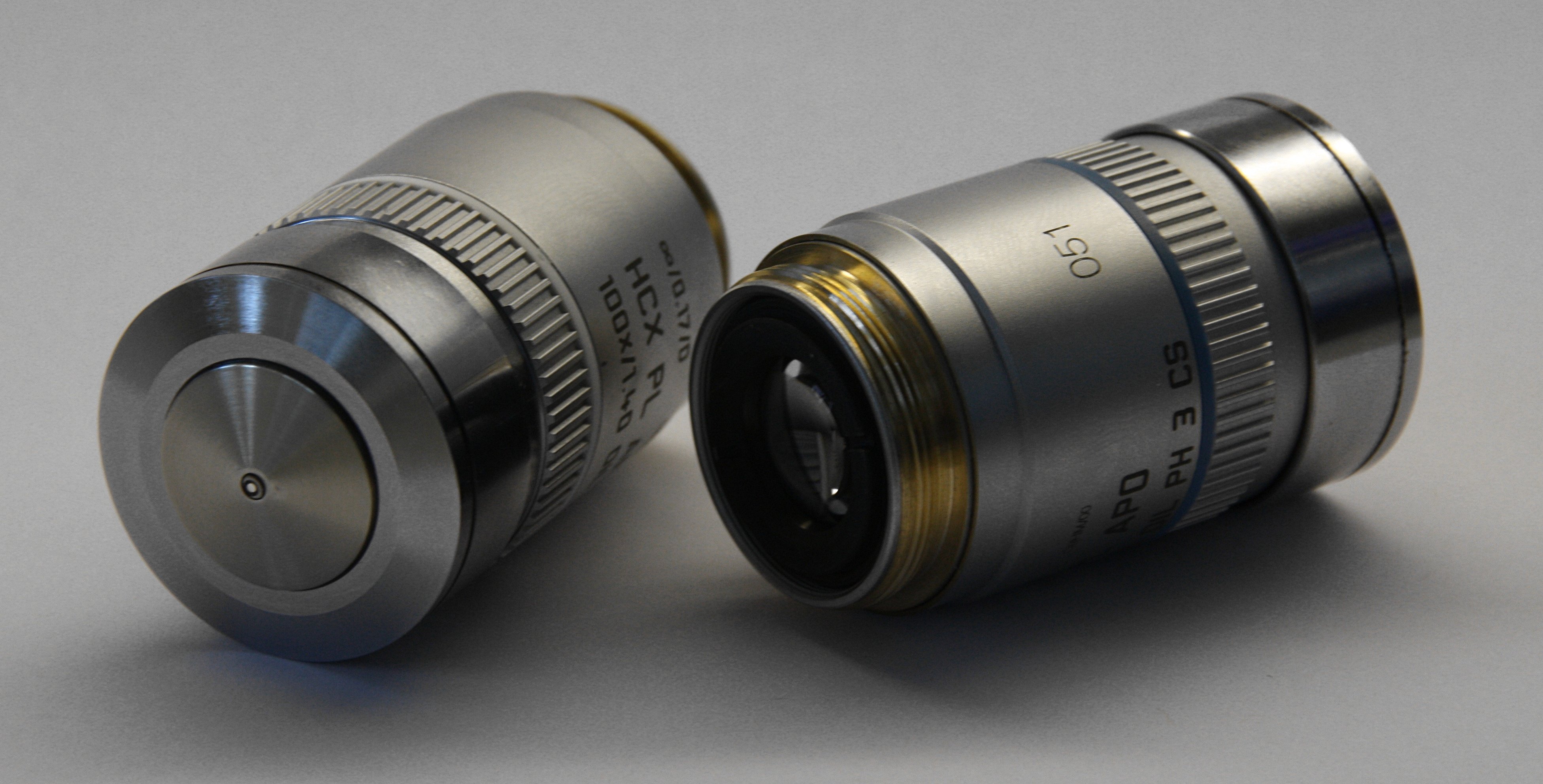 English: Two Leica oil-immersion epifluorescence m...
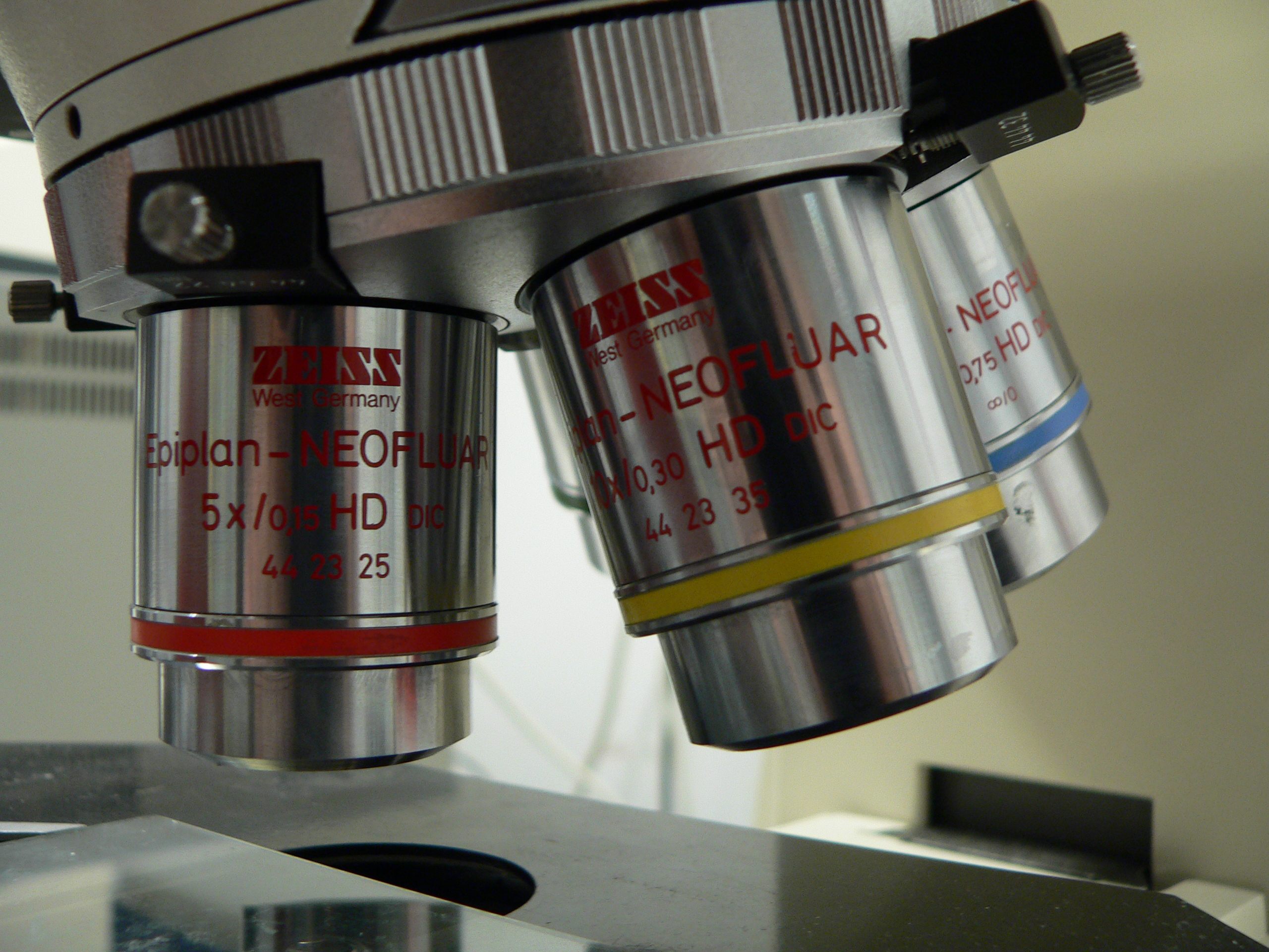 English: binocular microscope Français : Loupe bi...
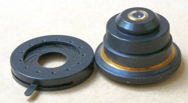 English: Microscope condenser lens.