Mitosis and Meiosis Mitosis: Cells can be divided in unicellular organisms or in multicellular organisms. DNA controls the cell division. Bacteria reproduce by a process called binary fission. Bacteria have one chromosome that's attached to the cell membrane. The chromosome replicates and the two copies separate as the cell grows. Over a period of time this one cell makes two cells. Eukaryotes do the process of mitosis. In mitosis, each daughter cell gets about half of the cytoplasm from the mother cell and one copy of the DNA.
Cells have to replicate the chromosomes so each daughter cell can have a set before cell division can occur. When the chromosomes have replicated and are ready to divide they consist of two identical halves called sister chromatids. A central region called the centromere joins the sister cromitids. Each individual chromosome is a long molecule of DNA and proteins. DNA makes up the genes and chromosomes that are made up of genes.
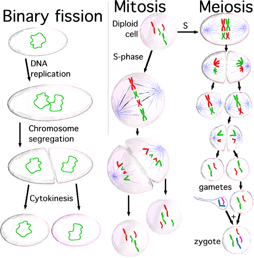 Three types of cell division: binary fision (takin...
 English: Cell into anaphase
 English: The stages of Mitosis: Interphase; early ...
In chromosomes some proteins turn off the genes that aren't needed in that cell.
In eukaryotes two basic types of cells occur in their bodies. Somatic cells are body cells that contain the same amount of chromosomes as each other inside the body of an organism. In somatic cells the number of chromosome is pretty consistent among the same type of species, but it varies from species to species. The chromosomes come in pairs, where one chromosome from each pair is from the mother and the other one is from the father. The other type of cell you would find in the eukaryotes is called gametes, also known as sex cells. Gametes consist of eggs in the females and sperm in the males. These reproductive cells have only has one set of chromosomes, which consist of one chromosome from each pair.
Mitosis is the process of chromosome division and cytokinesis is the process of cytoplasm division to enable it to form two cells. In almost all cells cytokinesis occurs with the last stage of mitosis. Centrioles are involved in chromosome division. Centrioles only occur in animal cells and contain nine sets of 3 microtubules. Each individual animal cell has a pair of centrioles that are located just outside the nucleus. The two centrioles in the pair are lined up at right angles to one another. The step before mitosis begins is where the centrioles replicate so the cell will have two sets of two as it starts to go into the process of mitosis.
The stages of mitosis include (interphase), prophase, metaphase, anaphase, and telophase. ~Interphase: Interphase may seem to be a stage kind of like a resting stage, but it's not. Cell growth, replication of the chromosomes, and many other kinds of activities and things go on in this phase. If the cell happens to be an animal cell, and it's near the end of interphase, the centrioles replicate so there are two pairs. The DNA strands, that make up the chromosome, now unwinds within the nucleus but don't look like real chromosomes. This genetic material is often known as chromatin.
~Prophase: The first division in the M phase of the cell division is called prophase. Prophase starts when the cyclin B/CDK2 complex (or kinase) reacts with the nucleic acid-protein complex, chromatin. When the two react, the kinase shortens the length of the chromatin to form pairs of sister chromosomes, called chromatids. During the time when this new reaction begins, DNA stops making certain RNA and the site where this RNA was produced the nucleus disappears. Next, a mitotic spindle mode of three different types of tiny tubule structure called microtubules. This occurs when micotubules, called astral fibers, come out of the top and bottom of the forming spindle. Then kinetochore MT connects the centromeres to the fibers. Finally, interpolar MT extends from the top or bottom to the middle.
The spindle is needed for three reasons: 1). To align chromosomes, 2). Pole movement, 3). Separate nuclei ~Metaphase: The next stage of mitosis is metaphase. When this stage begins, The nuclear envelope of the cell breaks down into vesicles and endoplasmic reticulum (ER). ER is a system of tubes -and sacs found in the cytoplasm of the eukaryotic cell. The newly formed mitotic spindle moves into nuclear region along with the vesicles. The chromatids attach to the spindle and move toward the center until they reach the midpoint. Then they align themselves to the opposite poles along the metaphase plate. The chromatids stay in the center because the poles act as magnets by applying equal force on either side of the chromosomes.
~Anaphase: The main part of anaphase is the separation of chromatids and the division of the centromeres. The separation begins as the microtubules pull on the kinetochase of each chromosome of a chromatid pair. These microtubules also work to push the poles of the mitotic spindle in opposite directions, further pulling the chromosomes apart. Anaphase ends when the new separated chromosomes converge at the poles of the spindle.
~Telophase: The final stage of mitosis is telophase. It's the formation of the two new cells. The beginning of telophase is the complete separation of the groups of chromosomes at each pole. These groups of chromosomes then form a nuclear envelope around them. In the final part of telophase the chromosomes return to their tangled, unorganized state. The cell splitting is completed after this.
~Cytokinesis: Cytokinesis is the process that takes place directly after mitosis. The final part of telophase in mitosis is included in cytokinesis. In animal cells, it is an indentation near the center of the cell groups and eventually pinches off because of a layer of actin and myosin. In plant cells, a cell plate that will eventually turn into cell walls forming between the dividing cells. The newly formed cells contain all the properties of complete, functioning cells.
Meiosis: ~Prophase 1: Prophase 1 is the first step in meiosis. Prophase 1 in meiosis is very similar to prophase of mitosis, but there are some differences. At the beginning of this stage, the cell chromosomes condense and become visible. In the S-phase of meiosis, each chromosome is replicated and in meiosis 1 they pair at centromeres to form bivalent. These homogenous chromosomes then pair one another to form synapses. Also, during this stage the nuclear membrane and the nucleolus dissolve.
~Metaphase 1: This next stage is called metaphase 1. This is the stage where the microtubules position the homogenous chromosome pairs, called tetrads, on the metaphase plate directly between both poles of the spindle.
~Anaphase 1: At the beginning of anaphase 1 the newly formed tetrads break down. Next, the synepsos break down and the microtubules pull the pairs of homogenous sister chromatids toward opposite sides of the cell. Each pole of the cell now has become haploid.
~Telophase 1: The last stage of meiosis 1 is telophase 1. By this stage the homogenous chromosomesare at opposite poles. The microtubules then disappear and nuclear envelopes may reform around the new cells.
~Cytokinesis: Cytokinesis in meiosis 1 is the same as in mitosis so there's no reason to point in writing it out again.
~Prophase 2: Prophase 2, the first step in the second meiotic division. It begins once more with the condensation of the chromatin. They then join again into sister chromatids. As in prophase 1, the nuclear envelope dissolves, and the microtubules form to pull the chromatids toward the metaphase plate at the center of the cell.
~Metaphase 2: Metaphase 2 begins when the chromatids are aligned along the metaphase plate. (Same as metaphase in mitosis.) ~Anaphase 2: In anaphase 2, the microtubules pull apart the sister chromatid pairs of their centromere. Then they move the split up pairs to opposite poles.
~Telophase 2: The final phase in meiosis is telophase 2. The single chromotids have reached the poles and the cell separates. New nuclear envelopes form around the chromatids. The result is four haploid cells.
~Comparing mitosis and meiosis Chromosome behavior 1. Mitosis: Homologous chromosomes are independent 2. Meiosis: Homologous chromosomes pair to form bivalents until anaphase 1.
Chromosome number (reduction in meiosis) 1. Mitosis- identical daughter cells 2. Meiosis-daughter cells haploid Genetic identity of progeny: 1. Mitosis- identical daughter cells 2. Meiosis- daughter cells have new assortment of parental chromosomes 3. Meiosis- chromatids not identical, crossing over
 Interphase nucleus of an HT1080 cell with part of ...
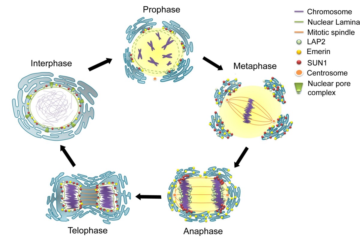 Nuclear envelope breakdown and reassembly in mitos...
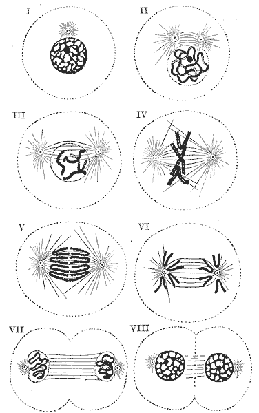 An illustration of the stages of mitosis in a huma...