Antibiotic action: Cellular structures and processes targeted by antibioticsBacteria are our friends and foes. They are everywhere , inside the body and outside. These smallest living organisms, very helpful for maintaining health and the environment . However they can be foes and actually threaten life by causing diseases. However , if bacteria became extinct eventually would we.
Antibiotics cure and prevent bacterial disease andl infections. Alexander Flemming , in 1928 , discovered penicillin , the first antibiotic. Eversince science has never surrendered. Different antibiotics are discovered to fight infections .
Bacteria are unicellular, prokaryotic organisms . Bacteria are round , spiral or rod shaped . Bacteria have a capsule for protection against drying out , cellwall which gives it shape and , a plasma membrane made of proteins and phospholipids allowing material exchange , nucleoid which contains DNA and ribosomes where translation occurs . Cellwall is made up of peptidoglycan , a feature unique to bacteria .
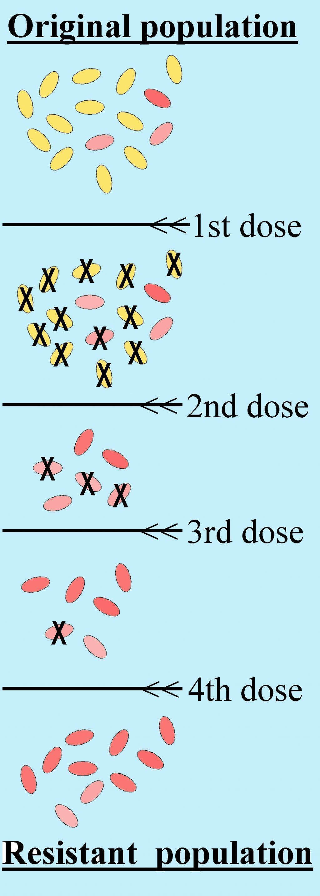 Schematic of the acquisition of antibiotic resista...
 English: example of selection for bacteria by anti...
 46:365 - Antibacterial Pinks
They are gram negative or positive . Metabolic activities of the bacterial cell differ significantly from those in the human cell .
Antibiotics are a selective poison disrupting specific functions in bacterial cell not in human cells They may be bacteriocidal which kill or bacteriostatic , which inhibit growth . Penicillin effects only gram+ bacteria but broad spectrum antibiotics work against both types . Antibiotic doses may cause fatal side-effects .
Antibiotics have various modes of action on a bacterial cell. Antibiotics target the sites and stages of cell wall synthesis . Some block DNA activity and cause DNA fragmentation . Consequently altering the structure of the template DNA and disrupting transcription. Many antibiotics work by targeting the ribosomes and RNA polymerase interferes with the synthesis of RNA, and consequently with protein synthesis. Some act by causing changes in the membrane. They replace...