QUEST TO KNOWING CELLS
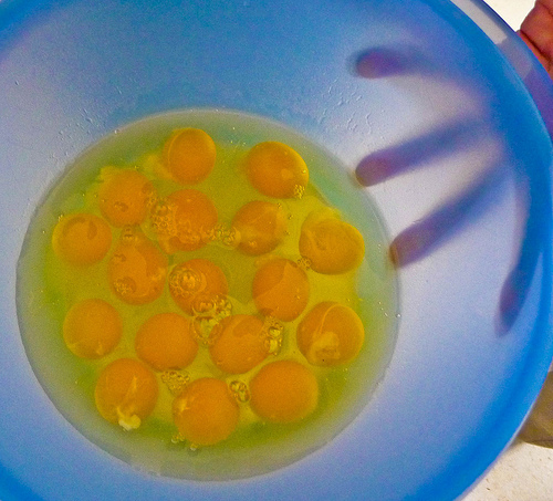
Stains are used to improve the contrast of the specimen so that the cells can be easily seen using the light microscope. Staining can damage the cells and possibly kill them; the nature morphology may be altered. The cheek cell should be scraped off gently to avoid damaging and crushing the plasma membrane and only two or three times to avoid injury to the cheeks. (Anonymous, 2011) Human cheek cells- no fixed shape, no cell wall, vacuoles absent, generally smaller in size, not arranged in a fixed pattern. Onion epidermal cells- fixed shape, cell wall, vacuoles present, generally bigger in size, arranged in a regular pattern. (Gan, 2009)
Cells must be discussed appropriately and accurately. The epidermal cells of onions and human cheek cells cannot be seen by the unaided eye but can be seen under light microscope. For total magnification of x100, the onion and cheek cells appear to be very small with the nucleus slightly visible.
 Cell biology
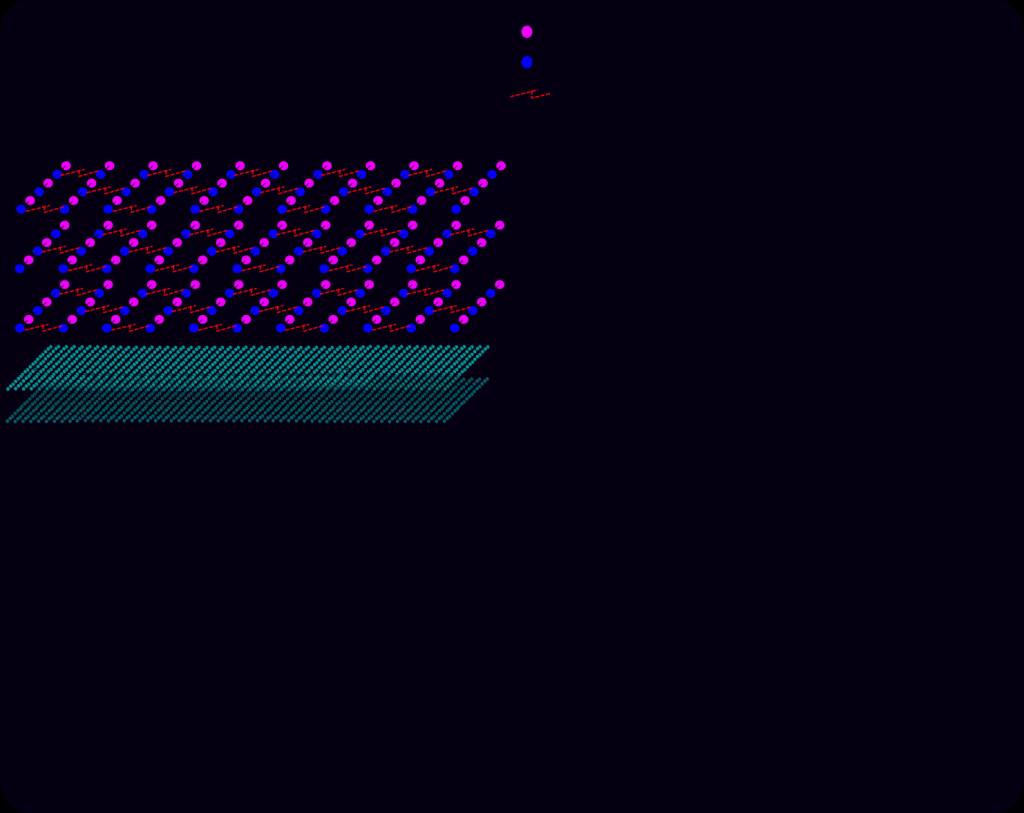 English: Schematic of gram-positive cell wall show...
 Cell Biology
As the magnification increases, the cells become clearer and the structure of the onion and cheek cells can be compared. Many cells appear quite transparent under microscope and are quite difficult to see. By staining the onion and cheek cells, a better contrast is provided so that the cells can be observed clearly and the shape, size and structure of the cells can be determined easily.
While observing the slide containing the onion cells, it is noticed that the onion cells have a regular shape. They are generally rectangular in shape as each cell is surrounded by a cell wall. The cell wall is made of cellulose and forms much of the supporting framework of the cell. Each cell also has a prominent nucleus, a large vacuole and the cytoplasm surrounding the vacuole. The nucleus is slightly pressed...